Health
How do I tell corneal dystrophy from an ulcer?
Dystrophy is in both eyes, painless and slow. An ulcer is in one eye, painful, red, and appears within hours. A stain test settles it in a minute.

Quick answer
How do I tell corneal dystrophy from an ulcer?
Dystrophy is in both eyes, painless and slow. An ulcer is in one eye, painful, red, and appears within hours. A stain test settles it in a minute.
Two facts separate this from everything else that makes a cornea cloudy: it is symmetrical, and the dog does not care. If either of those is untrue, you are probably not looking at dystrophy.
Owners who have noticed a whitish patch on the front of one or both eyes and are trying to judge whether it is an emergency, and working-dog owners who need to know whether it will affect the dog's job.
This guide is not medical advice. If your dog shows pain, sudden behavior change, or worsening symptoms, consult a licensed veterinarian.
Some links may be affiliate links. We may earn a commission at no extra cost to you. We only suggest products we believe are helpful for dog owners.
We research and check what we publish, but we do not warrant that any figure, date, price, or statement is accurate, complete, or current, and we accept no liability for any loss, cost, injury, or other harm arising from the use of this site. Use it as one reference among several. You remain responsible for your own decisions and for your dog's care - confirm anything that matters with a licensed veterinarian or another qualified professional before you act on it. Full disclaimer.
Corneal dystrophy is an inherited, non-inflammatory accumulation of fat or mineral within the clear front surface of the eye. It usually appears in both eyes in a similar position, is painless, and progresses slowly or not at all. Its main practical significance is that it can mask a genuinely dangerous corneal injury in a dog that works cover.
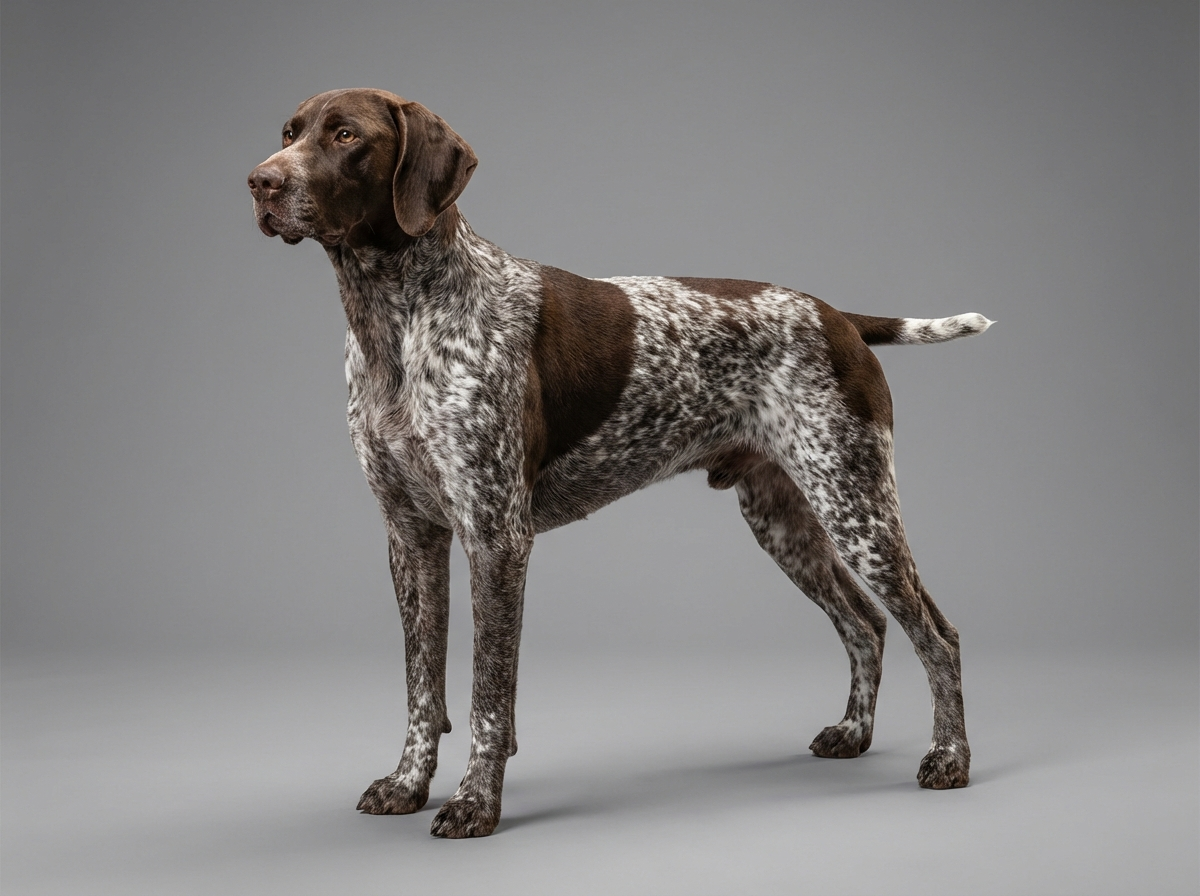
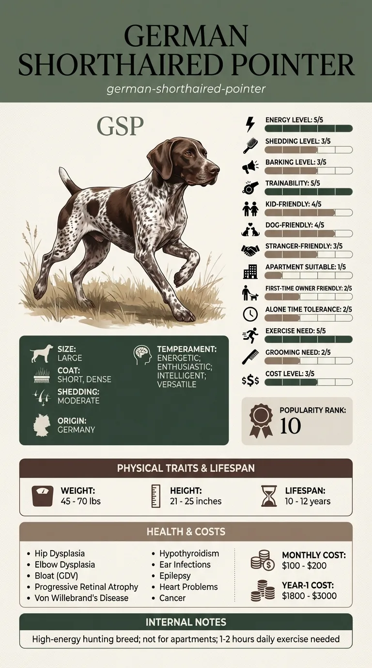
Corneal dystrophy is not listed among the German Shorthaired Pointer's documented health issues, which name progressive retinal atrophy as the breed's recognised eye problem. It occurs in a range of other breeds. A GSP owner is considerably more likely to be dealing with a corneal scar or ulcer from field work than with true inherited dystrophy.
The German Shorthaired Pointer has no documented predisposition to corneal dystrophy, and its recorded eye condition is retinal rather than corneal. Where the breed does contribute is to the differential. A dog developed in Germany as a versatile gundog, expected to quarter cover with its nose down, presents its corneas to seed heads, thorns and stems thousands of times a season. Scarring from healed injury looks very similar to inherited deposit, and distinguishing the two requires an examination rather than a photograph.
Environment does not cause dystrophy but shapes everything around it. Late-summer seeding grasses, cut stubble and dense brambles are the settings where corneal injury happens. Dry, windy conditions add grit. And a working routine that means an owner rarely looks closely at their dog's eyes in good light is the environmental factor most likely to let a real problem hide behind a known one.
Book an appointment to get the initial diagnosis confirmed with a fluorescein stain test, so you know what you are looking at rather than assuming. After that, go the same day for any of the following: an eye held shut or squinting, redness in the white of the eye, a new cloudy area in one eye only, a visible dent or dull spot on the surface, coloured or bloody discharge, or a dog pawing at its face. A corneal ulcer can deepen and perforate within days, and in a breed where the deposit is already familiar, the danger is dismissing a genuine injury as more of the same.
See all German Shorthaired Pointer health problems, which breeds are prone to corneal dystrophy, or the full German Shorthaired Pointer breed guide for temperament, exercise needs and ownership costs.
Diagnosis is a single appointment. After that, most dogs are simply monitored at annual health checks, with the deposit unchanged or very slowly enlarging over years. Expect no treatment and no restriction. Where a mineral deposit does break through and ulcerate, treatment usually resolves it over one to three weeks of medication.
A confirmed, benign diagnosis, a set of dated photographs, and an owner who has stopped worrying about the patch itself while becoming noticeably better at spotting the injuries this breed actually collects. The dog's work, vision and comfort are unchanged.
Both produce a cloudy cornea. One can be watched at a routine appointment; the other can cost an eye if left over a weekend. These are the features that separate them.
In a household pet, an owner who spots a painless white patch and mentions it at the next annual check has done everything necessary. In a German Shorthaired Pointer working cover several days a week, the same relaxed approach carries a real risk, and the reason is arithmetic rather than anatomy.
This is a breed that hunts with its head at ground level, at a gallop, through seeding grasses, stubble and brambles. Corneal injuries are an occupational hazard, and a dog with a pre-existing white patch on its cornea gives its owner a dangerous habit: seeing cloudiness and thinking that is just his eye. An ulcer developing on or beside an existing dystrophy is easy to overlook for exactly that reason, and a deep corneal ulcer can perforate.
The practical answer is to have the diagnosis confirmed once, properly, with a stain test and a look at both eyes, and then to photograph the patch so you have a reference. After that, any change in size, any redness, any squinting, or any new cloudiness in only one eye is a reason to be seen rather than a reason to shrug. The confirmed diagnosis is what makes it safe to relax about the thing you already know is there.

Hazel was six when a faint silvery haze appeared in the centre of both corneas, in mirror-image positions. She was untroubled by it, and the vet confirmed corneal dystrophy with a stain test that showed no ulceration. Her owner photographed both eyes and stopped thinking about it. Eighteen months later she came back from a hedgerow squinting on the right. Her owner's first thought was that the patch had got worse. Comparing the eye to the photograph on her phone showed something different: the left eye was unchanged, and the right had a new dull area beside the old haze, with a red rim around the white. It was a corneal ulcer from a thorn. Treated within a few hours it healed over ten days with drops and a buster collar. Her vet's remark was that dogs with known corneal deposits are the ones who most often arrive late, because their owners already have an explanation for cloudiness.
Key takeaway: The photograph did the work. Once you know your dog has a patch, the only way to keep noticing new problems is to have a record of exactly what the old one looked like.
The common form is not. Deposits sit within the layers of the cornea without inflaming it, and dogs behave completely normally. If your dog is squinting, tearing or rubbing its eye, something else is going on and it needs examining rather than watching.
Very unlikely. Most dystrophies are small, peripheral or faint enough that they interfere little with vision. A large central deposit can scatter light and reduce clarity, but complete vision loss from this condition is rare.
Generally it is monitored rather than treated, because the deposit itself causes no harm and interventions carry more risk than benefit. Where an underlying cause such as a lipid abnormality is found, treating that may stop progression. Surgical removal is reserved for unusual cases.
Corneal dystrophy is inherited in a number of breeds. It is not among the German Shorthaired Pointer's documented health issues, which list progressive retinal atrophy as the breed's recognised eye condition. Any GSP diagnosed with it is worth reporting to the breeder, since that is how breed data accumulates.
One particular form, where mineral deposits sit close to the surface, can break through and cause a painful ulcer. This is uncommon, but it is a reason to have any change in comfort taken seriously rather than assumed to be the same old patch.
No. A painless deposit that does not impair vision is no reason to change anything the dog does. What should change is the owner's eye-checking routine, because this breed's real corneal risk comes from the cover it runs through.
Dystrophy is in both eyes, painless and slow. An ulcer is in one eye, painful, red, and appears within hours. A stain test settles it in a minute.
Usually not. It is monitored. What needs treating is anything painful, red or one-sided that develops alongside it.
Photograph both eyes in daylight and keep the images dated. That reference is what lets you spot a genuine change later.
Very little. One diagnostic appointment with a stain test, then observation at routine visits. The cost that matters is treating any ulcer promptly.
A one-page prep sheet for this condition: the signs, the questions to ask, what to get priced. Unlocks here.
You get: Vet visit prep sheet
No spam. One-click unsubscribe. See our privacy policy.
Preview page 1 before you decide.
Early access list
The free prep sheet covers the same-day warning signs, a symptom log and the questions to ask. We are considering a fuller kit that adds a cost worksheet for the usual tests and treatments (US price ranges with their sources) and a plain-English guide to ways of covering the bill. It is not built yet; this list tells us whether it is worth building.
One email, no spam, unsubscribe anytime. Information only, not veterinary advice.