Health
What is IVDD Type I?
A sudden rupture of a prematurely calcified disc centre into the spinal canal, causing rapid severe compression. It is characteristic of short-legged chondrodystrophic breeds rather than pointers.

Quick answer
What is IVDD Type I?
A sudden rupture of a prematurely calcified disc centre into the spinal canal, causing rapid severe compression. It is characteristic of short-legged chondrodystrophic breeds rather than pointers.
The disc does not slide. It detonates. Everything about the emergency response follows from how quickly that damage is done.
Owners facing a sudden collapse in a large dog and needing to know how urgent it is, and anyone who has been given a grade number by a vet and wants to understand what it predicts.
This guide is not medical advice. If your dog shows pain, sudden behavior change, or worsening symptoms, consult a licensed veterinarian.
Some links may be affiliate links. We may earn a commission at no extra cost to you. We only suggest products we believe are helpful for dog owners.
We research and check what we publish, but we do not warrant that any figure, date, price, or statement is accurate, complete, or current, and we accept no liability for any loss, cost, injury, or other harm arising from the use of this site. Use it as one reference among several. You remain responsible for your own decisions and for your dog's care - confirm anything that matters with a licensed veterinarian or another qualified professional before you act on it. Full disclaimer.
Type I disc extrusion is an acute spinal cord injury caused by calcified disc material bursting into the spinal canal. It is a disease of chondrodystrophic breeds, and the German Shorthaired Pointer is not chondrodystrophic; spinal disease does not appear on its documented health list. The reason GSP owners still need this information is that acute hind-limb collapse from any cause is triaged the same way, on a five-point grading scale where the presence or absence of deep pain sensation carries most of the prognostic weight.
Type I extrusion is uncommon in German Shorthaired Pointers and should not be a routine worry for owners of the breed. It appears here because owners searching acute hind-limb weakness encounter Type I material written for dachshunds and need to know both that it probably does not apply and that the emergency response is the same regardless of which spinal problem is causing the collapse.
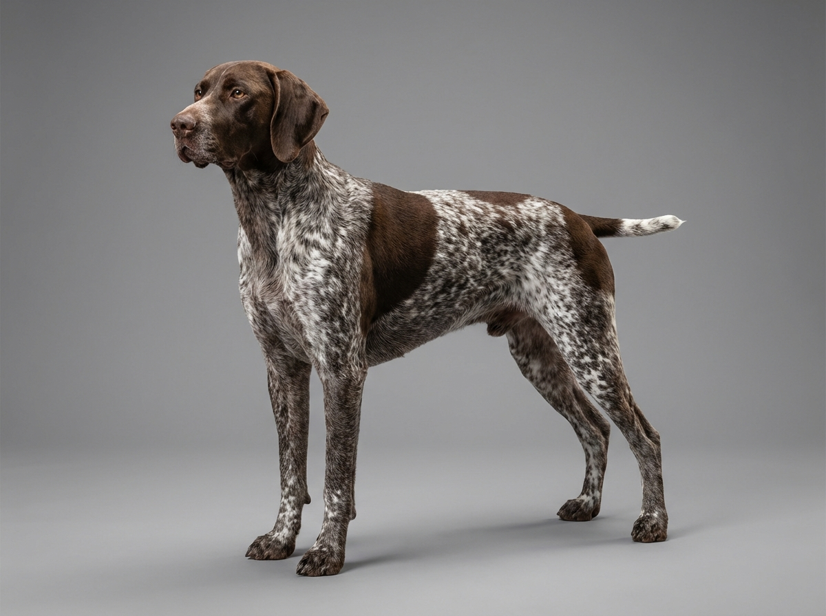
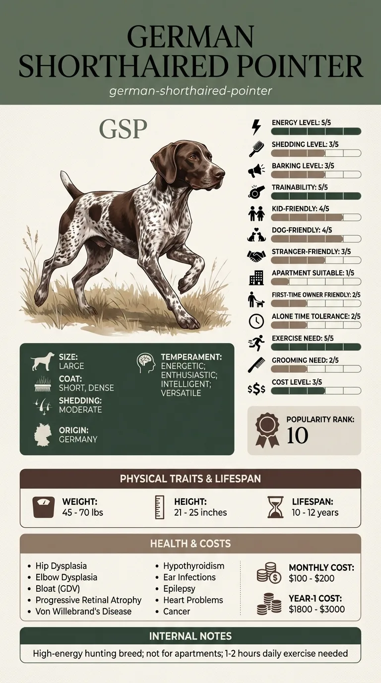
The defining breed fact is the absence of chondrodystrophy. A German Shorthaired Pointer has straight, correctly proportioned limbs and normal cartilage maturation, so its discs do not calcify early and are not primed to rupture explosively. Nothing on the breed's documented health list, which covers joints, gastric, ocular, bleeding, endocrine, ear, neurological seizure activity, cardiac and neoplastic disease, points toward disc pathology. What the breed does bring is exposure to force: a 45 to 70 pound dog rated five out of five for exercise need, working at speed through cover, occasionally meets an obstacle hard enough to damage a healthy spine.
For a non-predisposed breed the environmental contribution is almost the whole story. Vehicle strikes, collisions with gates and wire, falls from height and rough landings after jumps are the realistic mechanisms. Working terrain with ditches and hidden drops raises the odds. Household habits contribute at the margins, particularly repeated jumping down from high tailgates. None of this creates a chondrodystrophic disc; it simply applies enough force to injure a normal one.
Go immediately, without waiting for morning, if your dog cannot stand, cannot bear weight behind, is dragging its hind feet, has lost bladder control, or has sudden severe back or neck pain with any weakness. Support the spine flat during transport and phone ahead so the clinic is ready. Seek same-day assessment for hind-limb knuckling, crossing of the back legs, a newly wobbly gait, or acute pain without weakness, because grades can worsen within hours. Do not give human pain medication, several of which are toxic to dogs, and do not massage or manipulate a painful spine.
See all German Shorthaired Pointer health problems, which breeds are prone to intervertebral disc disease ivdd type i, or the full German Shorthaired Pointer breed guide for temperament, exercise needs and ownership costs.
Assessment and imaging happen within hours in a true emergency. Surgical cases typically involve several days of hospitalisation followed by six to twelve weeks of confinement and structured rehabilitation. Conservatively managed lower grades still require four to six weeks of genuine rest. Recovery of function in severe cases is measured in months, and improvement can continue well beyond the first few weeks.
At lower grades, success is a full return to normal function with a period of confinement behind you. At higher grades it is defined more modestly: independent walking even if imperfect, reliable bladder control, and a dog that is comfortable and engaged. Some dogs live well with permanent hind-end weakness and mobility support. Chasing a return to pre-injury field performance is not always the right target.
Neurologists grade spinal cord injury by function lost. The grade drives urgency and prognosis more than the imaging does.
The fibres carrying deep pain sensation are the most resilient in the spinal cord. They sit deep, they are widely distributed, and they survive insults that destroy motor pathways entirely.
That resilience is what makes them such a useful marker. If deep pain is still present, a great deal of cord tissue is still viable, and the outlook for recovering the ability to walk is considerably better. If it has gone, the injury has been severe enough to knock out the toughest fibres in the system.
This is why a veterinary examination includes firmly pinching a toe and watching not for a leg withdrawal, which is only a reflex, but for a conscious response: turning the head, vocalising, looking at the foot. Owners frequently mistake the reflex for sensation and offer reassurance the vet cannot use.
There is a further reason speed matters. A small proportion of severe cases develop a progressive softening of the cord that ascends along the spine and is not survivable. It is rare, and it is one of the reasons a dog that has lost hind-limb function is watched closely in hospital rather than sent home to see how things go.

Ansel had read about spinal injuries after a friend's dog was hurt, and kept a flat plywood offcut and an old blanket permanently in his truck. He felt slightly ridiculous about it for two years. Then his GSP, Perrin, ran into a wire strand at speed while working a hedge line and could not get up. Ansel slid the board under him, kept him straight, phoned ahead, and drove to a referral hospital ninety minutes away rather than the nearer local clinic. Perrin had deep pain sensation on arrival. Imaging showed a traumatic disc injury rather than the inherited kind. He had surgery that night, spent four days in hospital, and walked unaided again nine weeks later. The neurologist made a point of telling Ansel that arriving with the spine supported and going straight to a facility with an MRI had been worth more than anything else that happened before the anaesthetic.
Key takeaway: Preparation is the part of a spinal emergency you can control. Know how you will carry a 60 pound dog flat, and know which hospital has imaging, before the day you need both.
It is possible but atypical. Type I is driven by the chondrodystrophic cartilage trait that produces early disc calcification, and GSPs are normally proportioned dogs without that trait. Spinal disease does not appear on the breed's documented health list. Where a GSP does suffer an acute disc event, trauma is a more likely explanation than inherited degeneration.
Immediately, if there is any loss of hind-limb function. Do not wait overnight, do not wait for a regular appointment slot, and do not wait to see whether it improves. In severe grades the interval between injury and surgical decompression is one of the few variables that meaningfully changes the outcome.
Support the whole spine so it does not twist or sag. A rigid flat surface such as a board or a stretcher is ideal, with a second person if the dog is at the heavier end of the 45 to 70 pound range. Avoid lifting under the belly alone, which lets the spine drop, and keep the dog as still as possible in transit.
Plain radiographs cannot show the spinal cord and cannot reliably locate compression, though they help exclude fractures and some other conditions. Advanced imaging, usually MRI, is what identifies which disc space is involved and on which side, and surgery cannot sensibly be planned without it.
No. Lower grades with pain only or mild clumsiness are frequently managed conservatively with strict confinement and veterinary pain management, and many dogs do well. Surgery becomes the strong recommendation as function is lost, and the case for it strengthens sharply once a dog cannot walk.
Some do, and recovery can take months of intensive nursing and rehabilitation. Honesty matters here: outcomes at this grade are considerably less predictable than at lower grades, larger dogs are physically harder to nurse through it, and some never regain function. Your neurologist's assessment of your specific dog is worth more than any general figure.
A sudden rupture of a prematurely calcified disc centre into the spinal canal, causing rapid severe compression. It is characteristic of short-legged chondrodystrophic breeds rather than pointers.
Whether the dog can consciously feel a firm toe pinch. Those fibres are the most resilient in the cord, so their presence indicates substantial surviving tissue and a better outlook.
Yes, without qualification. Go to a veterinary clinic immediately, supporting the spine flat during transport, and do not wait to see whether it improves overnight.
On a rigid flat surface such as a board, with two people if possible, keeping the back straight rather than lifting under the belly and letting the spine sag.
Advanced imaging, surgery and hospitalisation together make this one of the largest single veterinary expenses a dog owner can face, before weeks of rehabilitation are added.
A one-page prep sheet for this condition: the signs, the questions to ask, what to get priced. Unlocks here.
You get: Vet visit prep sheet
No spam. One-click unsubscribe. See our privacy policy.
Preview page 1 before you decide.