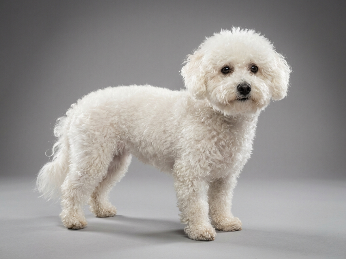
Health
How do I tell corneal cloudiness from a cataract?
Look from the side. A corneal deposit sits on the front dome of the eye; a cataract sits behind the pupil and changes visible extent as the pupil moves.

Quick answer
How do I tell corneal cloudiness from a cataract?
Look from the side. A corneal deposit sits on the front dome of the eye; a cataract sits behind the pupil and changes visible extent as the pupil moves.
Owners see a cloudy patch and think cataract, but a cataract sits deep inside the eye and a dystrophy sits on the front of it, and that difference changes almost everything about what happens next.
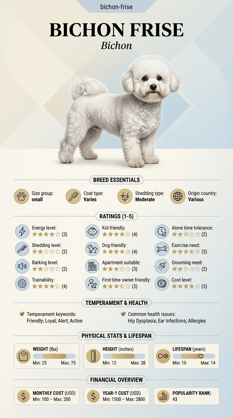
Owners who have spotted a pale mark on their Bichon's eye and are trying to work out whether it needs an appointment tomorrow or a mention at the next check. Also anyone who wants to know which cloudy eyes genuinely cannot wait.
This guide is not medical advice. If your dog shows pain, sudden behavior change, or worsening symptoms, consult a licensed veterinarian.
Some links may be affiliate links. We may earn a commission at no extra cost to you. We only suggest products we believe are helpful for dog owners.
We research and check what we publish, but we do not warrant that any figure, date, price, or statement is accurate, complete, or current, and we accept no liability for any loss, cost, injury, or other harm arising from the use of this site. Use it as one reference among several. You remain responsible for your own decisions and for your dog's care - confirm anything that matters with a licensed veterinarian or another qualified professional before you act on it. Full disclaimer.
Corneal dystrophy is an inherited accumulation of material, most often fats, within the layers of the clear front window of the eye. It appears as a well-defined whitish, silvery or crystalline patch, usually in both eyes and usually painless. Vision is rarely affected significantly. It is distinct from a cataract, which lies deeper in the eye, and from an ulcer, which is a painful loss of surface tissue and a genuine emergency.
Recognised in a range of breeds and generally regarded as a cosmetic finding in most affected dogs. Not documented in the Bichon Frise, whose eye-related entry on the health list is cataracts. Owners of this breed who notice cloudiness are statistically more likely to be looking at a lens change or, less often, at an acute surface problem than at a dystrophy.
The cornea's transparency depends on a highly ordered arrangement of collagen fibres and on the tissue being kept slightly dehydrated by a cell layer at its back. Dystrophy occurs when inherited differences in local metabolism allow material to be deposited between those fibres, scattering light. Because the underlying tendency is inherited, dystrophies cluster in particular breeds and often appear symmetrically in both eyes. The Bichon Frise carries no such documented tendency. Its inherited eye problem is cataract, a lens condition arising from an entirely different mechanism deeper inside the eye.
The environment does not cause dystrophy, but it does cause the acute corneal problems that owners must distinguish from it. A small dog at 9 to 12 inches moves through the world at the level of grass seeds, twigs, undergrowth and cat claws, which is why corneal scratches and ulcers are common in dogs of this height. Long facial hair, which this breed grows readily and which needs frequent grooming attention, can sweep across the eye surface and irritate it. Dusty homes, aerosols and dry heated air add ordinary irritation on top.
Mention a slowly appearing, painless, symmetrical cloudy patch at your next routine appointment, taking your photographs with you, and ask whether blood lipid testing is worthwhile. Seek same-day or emergency care for an eye that appeared cloudy suddenly, an eye held closed or squinting, obvious pain, redness, a watery or thick discharge, pawing or rubbing at the face, an eye that looks larger than the other, blood inside the eye, or any sudden change in your dog's vision. Corneal ulcers can deepen within a day and raised pressure inside the eye can destroy sight in hours, so eye pain is never a wait-and-see situation.
See all Bichon Frise health problems, which breeds are prone to corneal dystrophy, or the full Bichon Frise breed guide for temperament, exercise needs and ownership costs.
True dystrophies change slowly, and many stay essentially the same for years. Expect to be monitoring rather than treating, with a comparison at each annual examination. Where a cause such as abnormal blood lipids is identified and addressed, deposits may reduce over months, though they often persist. Acute problems run on a completely different clock: an ulcer treated promptly usually heals within one to two weeks, while one left untreated can deepen within days.
Success is a correctly categorised eye. That means a painless stable deposit that has been examined once, photographed periodically and otherwise left alone, in a dog whose sight is unaffected. It also means an owner confident enough to recognise the difference between that and a squinting, watery, suddenly cloudy eye, and who acts on the second within hours rather than filing it alongside the first.
Stand your Bichon somewhere with good natural light and look at the eye from the side rather than straight on. The cornea bulges forward as a dome; the lens sits behind the pupil, further back.
A corneal deposit sits on that forward dome. It usually appears as a sharply defined oval, arc or ring, often silvery or crystalline in appearance, frequently in a similar position in both eyes, and it does not move when the pupil changes size.
A cataract sits behind the pupil. It appears white or pearly, it is bounded by the edge of the pupil, and its visible extent changes as the pupil dilates in dim light and contracts in bright light.
The practical difference is that classic corneal dystrophy is usually painless, often non-progressive or very slowly progressive, and frequently needs no treatment at all beyond monitoring. Cataracts are on this breed's documented health list, can cause blindness and inflammation, and need veterinary assessment.
This is a triage exercise rather than a diagnosis. If you are unsure which you are looking at, that itself is a reason to book. What you are deciding is whether to phone today or mention it next month.

Most cloudiness is not urgent. A small number of presentations are, and they share a common feature: the dog is uncomfortable. Pain is the dividing line, and dogs show eye pain by squinting and rubbing rather than by crying.
If you think your Bichon has corneal dystrophy, the plan is three steps: write down what you have seen and when it started, book a veterinary appointment rather than waiting for the next flare, and take video of the behaviour or symptom before you go — the thing you are worried about rarely happens in the consulting room. Screening in the parents covers skin allergies, patellar luxation, bladder stones.
What the vet visit should produce is a diagnosis and a written plan, not just reassurance: what is being ruled out, what the monitoring interval is, and which signs mean you come back sooner. Ask what the treatment costs across a year rather than per visit, because that is the number that decides whether you are managing this condition or reacting to it.
Between appointments, keep a short log — dates, what you saw, what changed. It is the single most useful thing an owner brings to a follow-up, and for corneal dystrophy it is often what separates a clear pattern from a guess.
Iris noticed a small silvery oval on her Bichon Frise Monty's left cornea in March. It was sharply defined, he was not bothered by it, and there was a fainter one appearing on the right. She photographed both and raised it at his annual check in May. The vet examined the eyes, ran blood work that came back unremarkable, and recommended monitoring with repeat photographs. In August something else happened. Monty came back from a walk through long grass squinting hard on the right, with the eye streaming and his paw going repeatedly to his face. The eye had a dull patch that had not been there that morning. Iris phoned immediately rather than waiting for the annual pattern she had got used to. The examination found a corneal ulcer with a grass seed lodged under the upper lid. It was removed, treated, and healed in nine days. Monty still has his silvery ovals, unchanged three years later.
Key takeaway: The same dog can have cloudiness that needs nothing for years and cloudiness that needs a vet within hours, and the deciding question is always whether it came on suddenly and whether it hurts.
It is not on the breed's recognised health list, which covers skin allergies, patellar luxation, bladder stones, cataracts and dental disease. Corneal dystrophies are described in a number of breeds and can occur in individuals outside them. Cataracts are the eye problem genuinely associated with this breed.
Usually very little. The deposits are typically small, centrally or paracentrally placed, and dogs navigate around them without difficulty. Vision becomes a concern only if the affected area is unusually extensive, or if a different condition is present alongside it and has been overlooked.
In most cases no treatment is needed or recommended, and monitoring is the plan. Surgical removal of corneal tissue is reserved for particular circumstances because it can leave scarring, and scarring may obscure vision more than the original deposit did. An ophthalmologist is the right person to weigh that.
Photograph the eye head on, in consistent daylight, every few months, and keep the images dated. Comparing photographs over a year is far more reliable than comparing today's eye to a memory. Take the series to your annual appointment.
Almost certainly not. Dystrophies develop slowly over months and years. Sudden cloudiness points toward an ulcer, fluid in the cornea, or raised pressure inside the eye, and all of those are same-day problems. Sudden onset in an eye is one of the clearest signals to act quickly.
Fatty deposits in the cornea can sometimes accompany abnormal blood lipid levels or an underlying hormonal condition, so a vet may suggest blood tests alongside the eye examination. That is a sensible piece of thoroughness rather than a sign that something serious is suspected.
Look from the side. A corneal deposit sits on the front dome of the eye; a cataract sits behind the pupil and changes visible extent as the pupil moves.
Typically not. A painful cloudy eye is a different problem and needs same-day veterinary attention rather than monitoring.
Did it appear suddenly, and is the dog squinting or rubbing? Yes to either means today rather than next month.
Dated photographs head on in the same daylight every few months, kept together so a year of change is comparable at a glance.
Usually not. Most cases are monitored rather than treated, which keeps the cost to the routine examination you were having anyway.
A one-page prep sheet for this condition: the signs, the questions to ask, what to get priced. Unlocks here.
You get: Vet visit prep sheet
No spam. One-click unsubscribe. See our privacy policy.
Preview page 1 before you decide.