Health
What is corneal dystrophy in a dog?
An inherited deposition of material, usually lipid, within the layers of the cornea, appearing as a white or crystalline patch in both eyes and typically painless.

Quick answer
What is corneal dystrophy in a dog?
An inherited deposition of material, usually lipid, within the layers of the cornea, appearing as a white or crystalline patch in both eyes and typically painless.
Corneal dystrophy is one of the few eye findings in this breed that usually requires nothing at all. The reason it still matters is that it looks exactly like several things that are urgent.
Owners who have spotted a small white or silvery patch on the surface of one or both of their Chihuahua's eyes, and owners trying to work out whether a cloudy eye needs an appointment tomorrow or a drive tonight.
This guide is not medical advice. If your dog shows pain, sudden behavior change, or worsening symptoms, consult a licensed veterinarian.
Some links may be affiliate links. We may earn a commission at no extra cost to you. We only suggest products we believe are helpful for dog owners.
We research and check what we publish, but we do not warrant that any figure, date, price, or statement is accurate, complete, or current, and we accept no liability for any loss, cost, injury, or other harm arising from the use of this site. Use it as one reference among several. You remain responsible for your own decisions and for your dog's care - confirm anything that matters with a licensed veterinarian or another qualified professional before you act on it. Full disclaimer.
Corneal dystrophy is an inherited accumulation of material, most often lipid, within the layers of the cornea. It appears as a white, silvery or crystalline patch, characteristically in both eyes and roughly symmetrical, in an otherwise comfortable and quiet eye. It is generally not painful and rarely affects vision meaningfully. Its practical importance is that it resembles several urgent conditions, so the useful skill is distinguishing a quiet cloudy eye from an angry one.
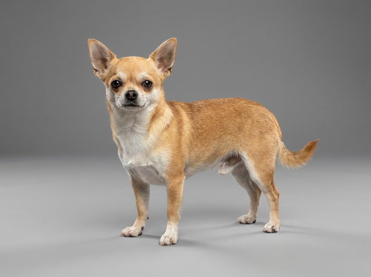
Corneal dystrophy is not among the Chihuahua's documented health problems, which include eye infections but not this specific finding. It occurs across many breeds and is more often a chance discovery at a routine appointment than a presenting complaint. Cloudy eyes in general are common in this breed, because prominent eyes in shallow orbits are more exposed to drying, irritation and injury than deep-set ones.
The mechanism of a true dystrophy is genetic and intrinsic to the cornea rather than related to Chihuahua anatomy, and it would be dishonest to invent a breed-specific cause. What the Chihuahua's build does supply is a reason this finding gets noticed and a reason it gets confused. The shortened muzzle of the apple-headed type produces shallow orbits and a prominent globe, so a larger proportion of the corneal surface is exposed and visible, and small changes are easier to see. That same exposure means the breed genuinely does acquire more corneal injuries, drying and infections, so any given cloudy patch in a Chihuahua has a reasonable chance of being something that matters.
Environment does not cause deposits inside the cornea, but it strongly influences the conditions that mimic them. Dry, heated indoor air, aerosols and scented products applied at floor level, dusty environments, and rough play with larger dogs all contribute to the surface irritation and ulceration that a worried owner may be looking at instead. Walks through dense undergrowth are a common source of corneal scratches in a dog whose eyes sit seven inches off the ground. A household that never looks at the eyes closely is the environment in which a real problem waits a week too long.
Go the same day for any cloudy eye that is also painful: squinting, held shut, red, weeping, or being rubbed on furniture. Go the same day too for an eye that has newly turned diffusely blue-grey, or where the pupil looks larger, smaller or a different shape from the other side, since raised pressure inside the eye damages vision within hours. Book a routine appointment for a stable, painless white patch present in both eyes, purely to have it identified and recorded. Never put leftover eye drops from a previous problem into a newly cloudy eye, since the wrong preparation on an ulcer makes it worse.
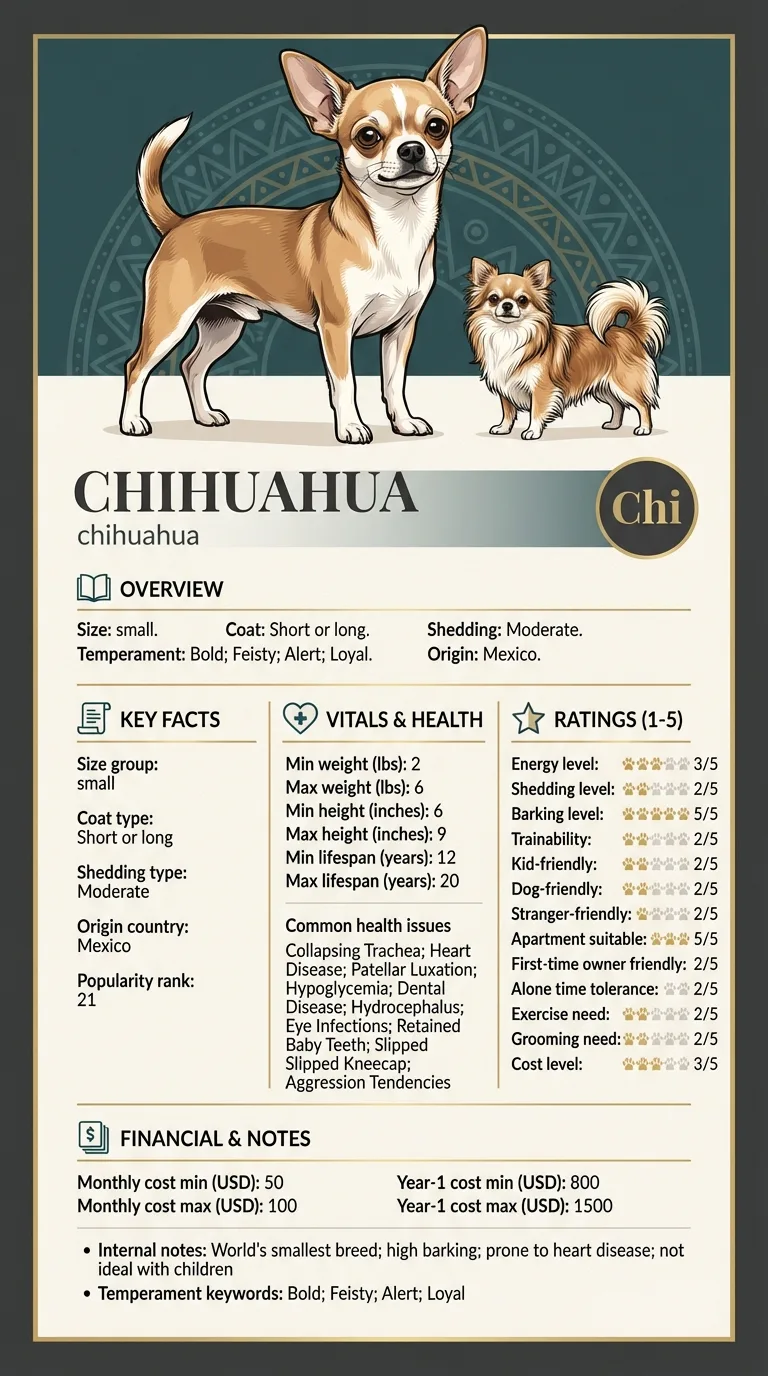
See all Chihuahua health problems, which breeds are prone to corneal dystrophy, or the full Chihuahua breed guide for temperament, exercise needs and ownership costs.
Corneal dystrophy typically appears in young to middle-aged adults and then changes very slowly, often remaining essentially static for years. Monitoring at routine annual appointments is usually sufficient. The innermost-layer form is different: it tends to progress gradually with increasing corneal swelling over months to years, and may eventually cause discomfort. Neither has a treatment timeline in the usual sense, because active treatment is generally not what is called for.
Success is an accurate label and a calm owner. That means a documented, photographed, dated finding that has been examined by a vet and identified as a dystrophy rather than an ulcer, an owner who checks both eyes daily and knows exactly which changes justify a same-day call, and a dog that is never treated for a problem it does not have. For most Chihuahuas with this finding, success is simply that nothing happens for the next ten years.
Corneal dystrophy is a genetically driven deposition of material within the cornea itself, most often lipid, appearing as a white, silvery or crystalline patch. Its signature is that it is bilateral and roughly symmetrical, often oval or ring-shaped, sits in the central or paracentral cornea, and is not painful. The eye around it is white and quiet, the dog does not squint, and nothing changes for months.
A corneal ulcer is also on the surface and can also look hazy, but it behaves completely differently. It hurts. The dog squints, the eye waters, the white of the eye goes red, and the dog paws at its face. Ulcers are diagnosed with a fluorescein stain that pools in the defect, and they need treatment quickly because a cornea this thin can perforate.
Corneal oedema is fluid within the cornea rather than deposits, and it produces a diffuse blue-grey cloudiness rather than a discrete white patch. It follows damage to the innermost corneal layer, and it can also indicate glaucoma, which is why a newly blue-looking eye is not something to watch over a weekend.
Behind all of this, cataracts and age-related nuclear sclerosis sit in the lens. The trick to placing them is depth: shine a light and see whether the cloudiness is on the front surface catching the light, or deep behind the pupil opening.

You cannot diagnose the layer at home, but you can reliably sort urgent from non-urgent, and that is the decision you actually need to make tonight.
If you think your Chi has corneal dystrophy, the plan is three steps: write down what you have seen and when it started, book a veterinary appointment rather than waiting for the next flare, and take video of the behaviour or symptom before you go — the thing you are worried about rarely happens in the consulting room. Screening in the parents covers collapsing trachea, heart disease, patellar luxation.
What the vet visit should produce is a diagnosis and a written plan, not just reassurance: what is being ruled out, what the monitoring interval is, and which signs mean you come back sooner. Ask what the treatment costs across a year rather than per visit, because that is the number that decides whether you are managing this condition or reacting to it.
Between appointments, keep a short log — dates, what you saw, what changed. It is the single most useful thing an owner brings to a follow-up, and for corneal dystrophy it is often what separates a clear pattern from a guess.
Sofia in Turin noticed a small silvery oval on the surface of her five-year-old Chihuahua Trufa's right eye, and a nearly identical one appeared on the left a fortnight later. Trufa was entirely comfortable, her eyes were white and wide open and she was not rubbing at anything. At the appointment the vet ran a fluorescein stain, which did not take up, confirming there was no ulcer, and identified bilateral corneal lipid deposits. No treatment was advised. Sofia was asked to photograph both eyes monthly in the same window light. Three years of photographs show almost no change. Where the routine did earn its keep was in the summer of the second year, when Trufa came in from the garden squinting hard with a watering, reddened left eye. Sofia knew instantly that this was not the patch, because the patch had never once made Trufa close her eye. It was a grass seed scratch, treated the same afternoon, healed in a week.
Key takeaway: The value of identifying a harmless finding is not the finding itself. It is that you now know exactly what your dog's normal cloudy eye looks like, so the day something genuinely wrong appears, you recognise it in seconds.
In its common forms, no. Lipid deposits within the corneal layers sit there without causing inflammation, and affected dogs behave completely normally. That is one of the diagnostic clues. The exception is a less common form affecting the innermost layer, which causes corneal swelling and can eventually produce small painful blisters on the surface, so a dystrophy that becomes painful needs reassessment.
Usually very little. The deposits are typically small relative to the whole cornea and the dog sees around them, in much the same way a small smudge on a windscreen does not stop you driving. Extensive deposits or the swelling associated with the innermost form can reduce clarity, but for most dogs the finding is cosmetic rather than functional.
Generally it is monitored rather than treated, because attempting to remove deposits from a cornea does more harm than the deposits do. Where an associated underlying condition exists, such as a lipid abnormality or a hormonal disease, treating that is worthwhile. Any treatment decision belongs to your vet, and surgical intervention is reserved for the small number of cases causing genuine problems.
By how the dog behaves rather than by how the eye looks. A scratch or ulcer is painful: squinting, watering, redness and pawing. Corneal dystrophy sits in a comfortable, white, wide-open eye. If your Chihuahua's eye has become cloudy and the dog is clearly uncomfortable, treat it as an ulcer until a vet says otherwise, because ulcers get worse quickly.
Corneal dystrophy is by definition a primary, usually inherited condition rather than one caused by injury or infection, and different breeds show different patterns of it. It commonly appears in young to middle-aged adults and affects both eyes. If you are breeding, it is a finding worth recording and discussing, since it is one of the things an ophthalmologic examination is designed to pick up.
Prominent, shallow-set eyes do raise this breed's overall ocular risk, and eye infections are on its documented health list. But that risk relates to exposure, drying and injury rather than to deposits within the cornea. What the prominent eye does mean is that a Chihuahua deserves a daily glance at both eyes, because it is more likely than most breeds to acquire something that genuinely needs attention.
An inherited deposition of material, usually lipid, within the layers of the cornea, appearing as a white or crystalline patch in both eyes and typically painless.
By pain. Dystrophy sits in a quiet, white, wide-open eye. An ulcer causes squinting, redness, watering and pawing at the face.
Usually not. It is monitored, since attempting to remove corneal deposits generally causes more trouble than the deposits themselves.
When it is painful, red, weeping, newly blue-grey, or the pupil looks different from the other side. Go the same day.
Surface cloudiness that catches the light sits in the cornea. Cloudiness visible deep behind the pupil opening sits in the lens.
A one-page prep sheet for this condition: the signs, the questions to ask, what to get priced. Unlocks here.
You get: Vet visit prep sheet
No spam. One-click unsubscribe. See our privacy policy.
Preview page 1 before you decide.