Autoimmune Disease in Papillons: When the Immune System Turns Inward
Quick answer
What is the fastest home check for autoimmune blood disease?
Lift the lip and part the belly hair. Pale or white gums point at red cell destruction; pinpoint red dots that do not fade under pressure point at platelet destruction. Either finding means a vet today.
Autoimmune disease is not an itchy problem. It is a pale-gums, pinpoint-bruising, sudden-collapse problem, and it moves on a scale of days.
Owners who have found something they cannot explain: a dog that has gone flat and pale overnight, tiny red dots on the belly skin, or crusting that has eaten away the smooth black surface of the nose.
Important reminder
This guide is not medical advice. If your dog shows pain, sudden behavior change, or worsening symptoms, consult a licensed veterinarian.
Some links may be affiliate links. We may earn a commission at no extra cost to you. We only suggest products we believe are helpful for dog owners.
We research and check what we publish, but we do not warrant that any figure, date, price, or statement is accurate, complete, or current, and we accept no liability for any loss, cost, injury, or other harm arising from the use of this site. Use it as one reference among several. You remain responsible for your own decisions and for your dog's care - confirm anything that matters with a licensed veterinarian or another qualified professional before you act on it. Full disclaimer.
What this problem looks like
Autoimmune disease is a group of conditions in which the immune system attacks the dog's own tissue. Depending on the target, a Papillon may present with sudden weakness and pale gums, unexplained bruising and pinpoint bleeding, slow weight gain and coat loss, crusting and pigment loss on the nose and pads, or a shifting lameness with fever. The blood forms are urgent; the endocrine and skin forms are chronic.
Common triggers
In most cases no trigger is ever identified, and the disease appears in a previously well dog
Underlying infection, particularly tick-borne disease in endemic areas
Certain medications, which can prompt an immune reaction against blood cells
Cancer elsewhere in the body driving a secondary immune-mediated process
Ultraviolet exposure, which can worsen established autoimmune skin lesions on the nose
Uncommon in dogs generally and not a recognised feature of this breed. Most Papillon owners will never encounter it. It earns a place on any health list because the presentation of the blood forms is dramatic, the window for treatment is short, and the early signs are things a person can check at home in seconds.
Why this happens
Breed factors
There is no established genetic predisposition to immune-mediated disease in Papillons, and the breed does not sit in the group vets watch for it. What the breed does contribute is visibility. Fine pigmented skin under a single silky coat means bruising and petechiae can be seen once the hair on the abdomen is parted, and the breed's characteristic alert, confident manner means a dog that has gone unusually flat is a change an owner notices early.
Environment factors
Environment plays a smaller part here than in allergic disease. The clearest environmental link is infection: tick-borne organisms in some regions can set off secondary immune destruction of blood cells, which makes tick prevention relevant beyond the parasites themselves. Sunlight worsens established autoimmune lesions on a depigmented nose, so a dog with that diagnosis needs shade rather than midday walks.
What you can do at home (once it is not an emergency)
When to talk to your vet
Go the same day, or out of hours, for pale, white or yellow gums; pinpoint red spots or unexplained bruising anywhere; blood in urine, stool or vomit; sudden weakness, breathlessness at rest or collapse. Immune-mediated destruction of red cells or platelets is a genuine emergency and dogs deteriorate over days, not weeks. Book a routine appointment for gradual weight gain with a thinning or dull coat, for crusting or loss of pigment at the nose, ear edges or footpads, and for lameness that moves between legs, especially with a fever or reluctance to move.
At home, alongside your vet's plan
Learn a thirty-second weekly check: gum colour, gum surface for pinpoint dots, and the thin skin of the belly and inner thighs parted with your fingers.
Photograph your dog's nose and ear margins while they are healthy, so any later change in texture or pigment can be compared rather than debated.
Keep tick prevention running through the whole season if you live where tick-borne disease occurs.
Note energy levels in a diary rather than trusting memory, since the slow forms are recognised by trend rather than by any single bad day.
If your dog is already on immune-suppressing treatment, keep a simple temperature log, because fever is the earliest sign of the infections that treatment invites.
Immune-mediated blood disease is fought over days. Expect several days of hospitalisation, then weeks of frequent blood tests as treatment is tapered, and a first assessment of whether the disease is controlled at around three to six months. Thyroid autoimmunity is different: replacement is started, a blood test at six to eight weeks confirms the level, and the dog is normal thereafter. Skin forms typically take two to three months to bring under control.
For blood disease, success is a stable red cell or platelet count on the lowest dose of medication that holds it, with a dog that is bright, eating and off the emergency list. For thyroid disease, success is a Papillon whose coat and energy come back and whose owner forgets there was ever a problem. For skin disease, it is intact pads and a nose that stops cracking, accepted alongside some residual pigment loss.
The families of autoimmune disease that affect dogs
Blood is the most urgent group. In immune-mediated haemolytic anaemia the body destroys its own red cells, leaving a dog weak, breathless and pale, sometimes with yellow-tinged gums or eye whites. In immune-mediated thrombocytopenia the target is platelets, and the sign is bleeding: pinpoint red dots on the belly and gums, bruising with no injury, or blood in urine or stool. Both can become life-threatening within days.
Endocrine autoimmunity is slower and quieter. Immune destruction of the thyroid gland produces months of gradual weight gain, low energy and coat change before anyone suspects a disease.
Skin autoimmunity, the pemphigus group, attacks the junctions holding skin cells together. The classic pattern is crusting and depigmentation at the nose, the ear margins and the footpads, rather than itch spread across the body.
Joint autoimmunity produces a dog that shifts lameness from leg to leg, often with a fever and a stiff, reluctant walk.
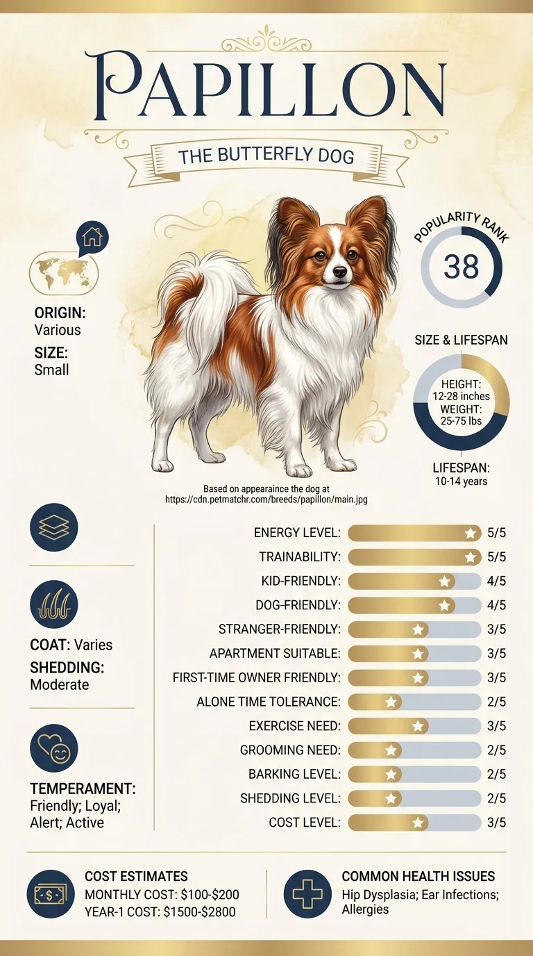
Autoimmune Disease in Papillons - The families of autoimmune disease
Where the Papillon actually sits
The breed's documented health list runs to patellar luxation, progressive retinal atrophy, dental disease, an open fontanel and a collapsing trachea. Autoimmune disease is not on it, and Papillons are not a breed vets associate with immune-mediated illness the way they do some spaniels, poodles and Old English Sheepdogs.
That is genuinely reassuring, but it changes only the odds, not the response. Any individual dog of any breed can develop these conditions, and the signs are the same whatever the pedigree.
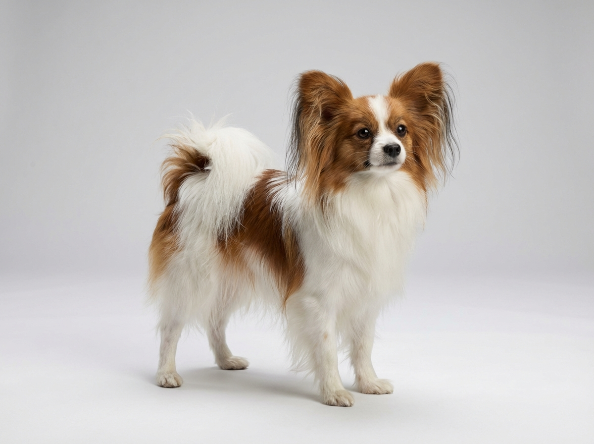
One practical Papillon detail is worth knowing. A long, silky single coat with fine pigmented skin makes pinpoint haemorrhages relatively easy to see once you know to part the hair on the belly and the inner thighs, where the coat is thinnest. Pale gums are also easy to assess in this breed, since Papillons tolerate having their lips lifted if the handling has ever been rewarded. Both checks take ten seconds and are the two most useful things a worried owner can do at home.
The dots Ruth found on a Sunday
Ruth was brushing the belly of her six-year-old Papillon, Wren, when she noticed a scatter of tiny red dots across the pink skin near the groin. Wren was behaving normally and eating supper.
Ruth had read once that spots which do not fade under pressure are not bruises. She pressed one with her thumbnail, saw it stay, and rang the out-of-hours service rather than waiting for Monday.
Wren's platelet count came back severely low. She was admitted that night and started on treatment before she had bled anywhere that mattered. Six months on she is on a small maintenance dose with blood tests every eight weeks, and her vet has said more than once that the Sunday phone call is the reason the story reads the way it does.
Key takeaway: The dots are not dramatic and the dog does not look ill. That combination is exactly why immune-mediated platelet disease is caught late, and why a weekly belly check is worth the ten seconds.
Frequently asked questions
Are Papillons prone to autoimmune disease?
Not notably. The breed's documented problems are orthopaedic, ocular, dental, skeletal and respiratory, and immune-mediated illness is not among them. Individual dogs still develop these conditions, so the signs are worth knowing regardless of the breed odds.
How is autoimmune disease different from allergy?
Allergy is the immune system overreacting to something external and mostly causes itch and inflammation. Autoimmune disease is the immune system destroying the dog's own tissue and mostly causes anaemia, bleeding, organ dysfunction or structural skin damage. The treatments are related but the urgency is completely different.
What are pinpoint red dots on my dog's belly?
Those are petechiae, small haemorrhages under the skin, and they are one of the few findings that should send you to a vet the same day. They usually indicate a platelet problem rather than a bruise, and they do not blanch when you press them.
Can vaccinations trigger autoimmune disease?
A temporal association has been described in some cases, but a causal link is not established and the great majority of autoimmune cases have no vaccine history behind them. Discuss the schedule with your vet if your dog has had an immune-mediated illness; do not stop core protection on your own.
Is treatment lifelong?
Often, and it usually involves medication that suppresses the immune system, with regular blood monitoring. Some dogs are eventually weaned down to a low maintenance dose or off entirely, while others relapse when treatment is reduced. Autoimmune thyroid destruction requires lifelong replacement rather than suppression.
What is the outlook?
It depends heavily on which system is attacked. Autoimmune thyroid disease has an excellent outlook once treated. Skin forms are usually controllable over years. Red cell and platelet destruction are far more serious and carry real mortality in the first weeks, which is exactly why speed matters.
What is the fastest home check for autoimmune blood disease?
Lift the lip and part the belly hair. Pale or white gums point at red cell destruction; pinpoint red dots that do not fade under pressure point at platelet destruction. Either finding means a vet today.
Health
Does crusting on the nose mean autoimmune skin disease?
Not necessarily, but it deserves investigation. Loss of the smooth cobblestone texture and pigment on the nose, with crusting at the ear edges or footpads, is the pattern that distinguishes it from ordinary dryness or sunburn.
Costs
What does treatment cost?
Initial hospitalisation for an immune-mediated blood disease commonly runs into thousands, and transfusion pushes it higher. Long-term medication and monitoring then add ongoing monthly cost well above this breed's usual eighty to one hundred and fifty dollar range.
Living
How do I care for an immunosuppressed Papillon at home?
Keep the dog away from other dogs' faeces and standing water, avoid busy dog parks, keep bedding clean and dry, and report any fever, cough or new lethargy quickly, since infection is the main risk of the treatment itself.
Training
Can training help during recovery?
Only indirectly, but it matters. A Papillon taught to accept gum checks, leg handling and being still for blood sampling makes months of monitoring far less stressful, and this breed learns cooperative-care routines quickly.
Related DogBreedCompass guides
immune-mediated thyroiditis in Papillons – Autoimmune destruction of the thyroid is the slow, common form and is treated very differently.
Papillon allergies explained – Allergy is the condition most often confused with autoimmunity by owners.
von Willebrand's disease in Papillons – Bleeding signs can also come from an inherited clotting disorder rather than an immune one.
skin infections in Papillons – Autoimmune skin disease has to be separated from ordinary bacterial skin infection.