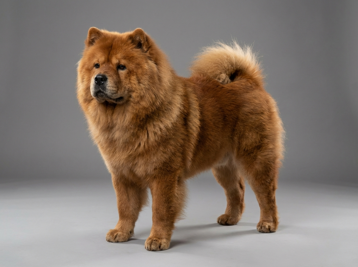
Health
How do I tell surface cloudiness from a cataract?
Look from the side. A surface opacity shifts with the curve of the cornea; a cataract sits behind the pupil and does not.

Quick answer
How do I tell surface cloudiness from a cataract?
Look from the side. A surface opacity shifts with the curve of the cornea; a cataract sits behind the pupil and does not.
Two questions sort almost every cloudy eye. Is the cloudiness on the surface or behind the pupil? And does the eye hurt? A painless, symmetrical, superficial haze is a very different situation from a single eye held half shut - and in a breed with documented eyelid disorders, the second is the more likely story.
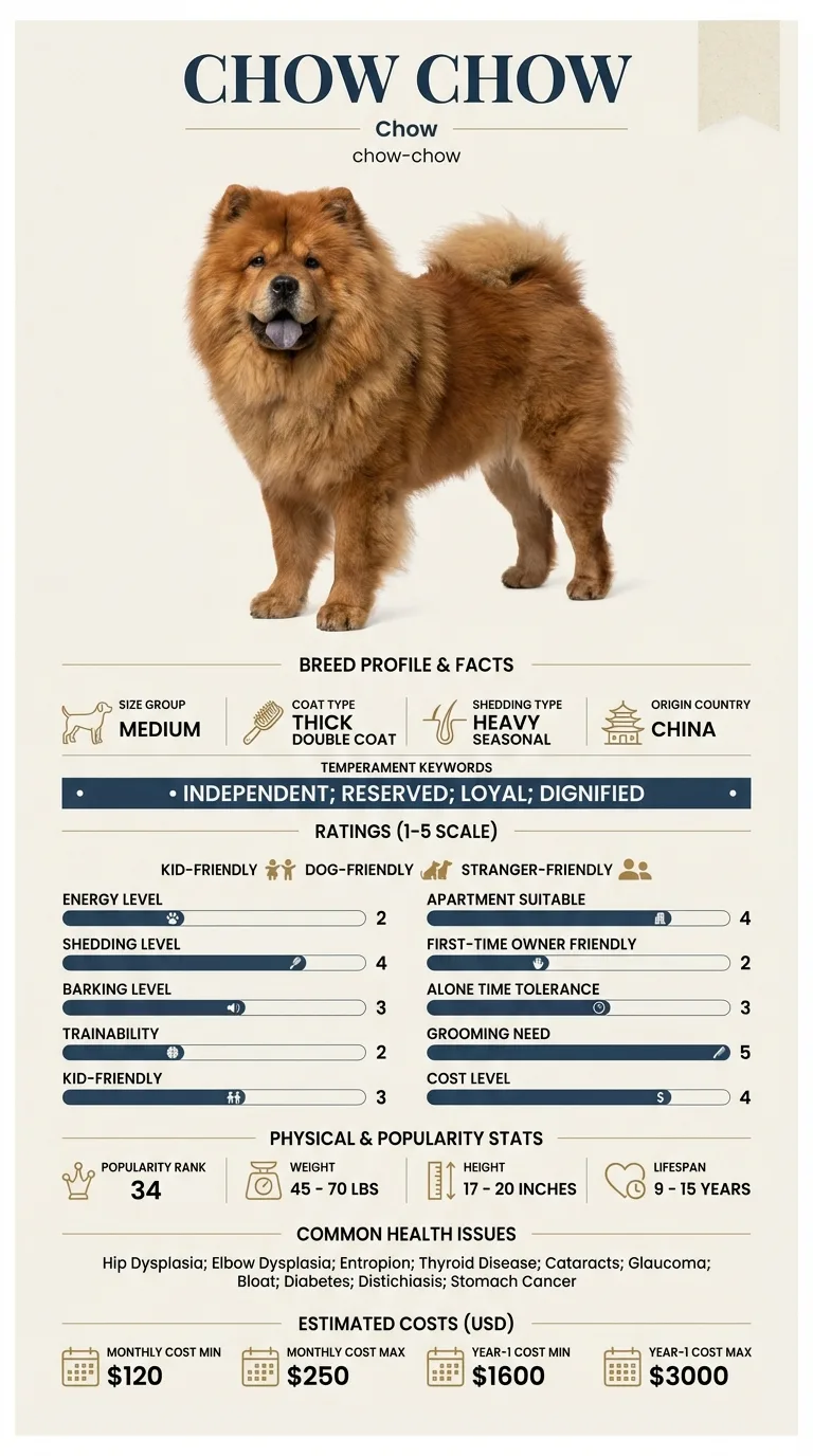
Owners who have spotted something on the eye and are trying to work out how urgently to act. It is particularly relevant for Chow owners, because this breed's documented entropion and distichiasis mean that hair or lashes rubbing the cornea is a realistic and painful alternative explanation.
This guide is not medical advice. If your dog shows pain, sudden behavior change, or worsening symptoms, consult a licensed veterinarian.
Some links may be affiliate links. We may earn a commission at no extra cost to you. We only suggest products we believe are helpful for dog owners.
We research and check what we publish, but we do not warrant that any figure, date, price, or statement is accurate, complete, or current, and we accept no liability for any loss, cost, injury, or other harm arising from the use of this site. Use it as one reference among several. You remain responsible for your own decisions and for your dog's care - confirm anything that matters with a licensed veterinarian or another qualified professional before you act on it. Full disclaimer.
Corneal dystrophy is deposition of fat or mineral within the clear surface of the eye, producing a usually symmetrical, usually painless white or grey haze. It is not documented in the Chow Chow. Its practical value for owners of this breed is as a comparison point: knowing what harmless cloudiness looks like makes it easier to recognise the painful, one-sided cloudiness that this breed's documented eyelid conditions actually cause.
Uncommon in this breed, which has no documented association with it. What Chow owners do encounter reasonably often is corneal damage secondary to entropion and distichiasis, both of which appear on the breed's documented condition list. In practice, then, a cloudy cornea in a Chow is more likely to be a consequence of the lids than a primary corneal disease.
The Chow Chow's eye risk comes from the lids rather than the cornea itself. Entropion, in which the lid margin rolls inward so that hair contacts the eyeball, and distichiasis, in which extra lashes emerge from the lid margin, are both documented in this breed. Each produces continuous mechanical abrasion of the cornea, and chronic abrasion leads to cloudiness, scarring and pigment deposition that can look superficially like a dystrophy. The breed's deep-set eyes under a heavy brow, surrounded by dense coat, also mean debris and hair are held close to the eye and that early changes are difficult for an owner to see.
Coat around the face is the most direct factor, since long hair over the eyes carries dust and debris onto the surface. Dogs that run through undergrowth or swim in untreated water pick up mechanical and infectious insults. Dry, dusty or heavily air-conditioned indoor environments reduce tear film quality. And any environment where a dog rubs its face repeatedly - usually because of an existing irritation - accelerates surface damage substantially.
Book a routine appointment for a painless, stable haze in a comfortable dog, and ask for a fluorescein stain so the question is settled properly. Go the same day for a painful eye: one held shut, watering, red, or being rubbed; sudden cloudiness in one eye; a visible defect or crater on the surface; or an eye that appears larger or is bulging. Acute corneal ulceration can perforate, and raised pressure inside the eye destroys vision within a day, so neither waits. Do not put any leftover or human eye product into a painful eye.
See all Chow Chow health problems, which breeds are prone to corneal dystrophy, or the full Chow Chow breed guide for temperament, exercise needs and ownership costs.
True dystrophy changes over years and is generally monitored at routine appointments rather than treated. Corneal ulcers heal within days to a couple of weeks with appropriate treatment, though deep or complicated ones take longer and may need surgery. Where an eyelid abnormality is the cause, corrective surgery resolves the source and the cornea then heals over the following weeks, sometimes leaving a permanent scar.
For dystrophy, success is a stable finding that never troubles the dog and is simply noted at each check-up. For irritation-driven cloudiness, success is the lids corrected, the cornea healed and no recurrence, accepting that a residual scar may remain. In both cases the real win is having a correct diagnosis early, because the two demand opposite levels of urgency.
Corneal dystrophy sits within the thickness of the cornea itself. It typically appears as a fairly well-defined oval or circular haze near the centre of the eye, it is usually present in both eyes in a roughly mirrored position, and the dog behaves entirely normally. It develops slowly, often over years.
Corneal oedema or ulceration also sits on the surface, but looks and behaves differently. It tends to be a diffuse blue-grey haze rather than a crisp white patch, it is frequently in one eye only, and the dog is uncomfortable - squinting, watering, rubbing at the face, avoiding light. This is the category that includes damage from an inward-rolling eyelid or an abnormal lash, both documented in the Chow Chow, and it needs prompt veterinary attention.
Cataract is not on the surface at all. It sits behind the pupil, inside the eye, and looks like a change in the colour of the pupil itself rather than a mark on the window. Cataracts are also documented in this breed.
Distinguishing surface from depth at home is easier than it sounds. Look at the eye from the side rather than straight on. A surface opacity moves with the curve of the cornea as your viewpoint changes; something behind the pupil stays put.

A painless cloudy eye can wait for a routine appointment. A painful one cannot, because the breed's documented eyelid problems can damage a cornea quickly and permanently.
If you think your Chow has corneal dystrophy, the plan is three steps: write down what you have seen and when it started, book a veterinary appointment rather than waiting for the next flare, and take video of the behaviour or symptom before you go — the thing you are worried about rarely happens in the consulting room. Screening in the parents covers hip dysplasia, elbow dysplasia, entropion.
What the vet visit should produce is a diagnosis and a written plan, not just reassurance: what is being ruled out, what the monitoring interval is, and which signs mean you come back sooner. Ask what the treatment costs across a year rather than per visit, because that is the number that decides whether you are managing this condition or reacting to it.
Between appointments, keep a short log — dates, what you saw, what changed. It is the single most useful thing an owner brings to a follow-up, and for corneal dystrophy it is often what separates a clear pattern from a guess.
Freya's owner, Malachy, noticed a greyish patch on her left eye and, having read about corneal dystrophy, was reassured to find that it was described as harmless. He nearly left it until her booster was due three months later. What changed his mind was a detail from the same reading: dystrophy is usually symmetrical. Freya's right eye was completely clear. He also realised, once he started paying attention, that she was blinking more on the left and had a damp patch in the coat below that eye. The fluorescein stain lit up a small ulcer. The cause was two fine extra lashes growing from the inside of her left upper lid, which had been rubbing the surface every time she blinked. They were treated, the ulcer healed in nine days, and Malachy was told the eye would probably keep a faint scar.
Key takeaway: Symmetry and comfort are the two things to check before reassuring yourself. A cloudy patch in one eye only, in a dog that is blinking or watering on that side, is not the harmless diagnosis - and in a breed with documented lash and lid problems, it usually has a correctable cause sitting right above the eye.
The breed's documented eye conditions are entropion, distichiasis, cataracts and glaucoma. Corneal dystrophy is not among them, and it is more strongly linked with certain other breeds. If your Chow has a cloudy cornea, the documented lid conditions are the more probable underlying explanation and should be excluded first.
Frequently not. Where it is genuinely dystrophy, does not affect vision and causes no discomfort, the usual approach is monitoring. Treatment becomes relevant if the deposits break through the surface and cause ulceration, or if vision is meaningfully affected, both of which are less common.
Usually with a fluorescein stain, which is a dye that adheres to damaged corneal surface and shows up under blue light. A stain-positive eye has an ulcer; a stain-negative white patch is more likely a dystrophic deposit or scar. It is a quick, inexpensive test and it settles the question.
Yes, and this is the key point for a Chow. Entropion rolls the lid inward so that hair contacts the cornea, and distichiasis produces extra lashes from the lid margin. Both abrade the surface and cause cloudiness, scarring and pigment deposition over time, and both are correctable surgically.
No. Some preparations, particularly steroid-containing ones, can seriously worsen an ulcerated cornea and delay healing. Never apply a leftover eye medication to a new problem without a vet examining the eye and confirming the cornea is intact.
True dystrophy usually progresses very slowly and many dogs live with it uneventfully. What can get worse quickly is anything caused by ongoing mechanical irritation, which is why establishing whether lashes or a rolled lid are involved is the more urgent question in this breed.
Look from the side. A surface opacity shifts with the curve of the cornea; a cataract sits behind the pupil and does not.
If it is painful, red, watering or held shut, yes. A painless symmetrical haze in a comfortable dog can wait for a routine appointment.
Because entropion and distichiasis are documented in the breed, so a damaged cornea has an obvious and correctable cause worth finding.
A fluorescein stain is quick and cheap, and it is what separates a harmless deposit from an ulcer needing treatment.
A one-page prep sheet for this condition: the signs, the questions to ask, what to get priced. Unlocks here.
You get: Vet visit prep sheet
No spam. One-click unsubscribe. See our privacy policy.
Preview page 1 before you decide.