Health
What does cherry eye look like?
A smooth, rounded pink or red mass at the inner corner of the eye, usually appearing suddenly rather than developing gradually.

Quick answer
What does cherry eye look like?
A smooth, rounded pink or red mass at the inner corner of the eye, usually appearing suddenly rather than developing gradually.
The single most important thing to know is what must not happen. That gland is not spare tissue - removing it, which used to be routine practice, permanently reduces tear production and sets the dog up for a lifetime of dry eye. If anyone offers to simply take it out, ask about replacement instead.
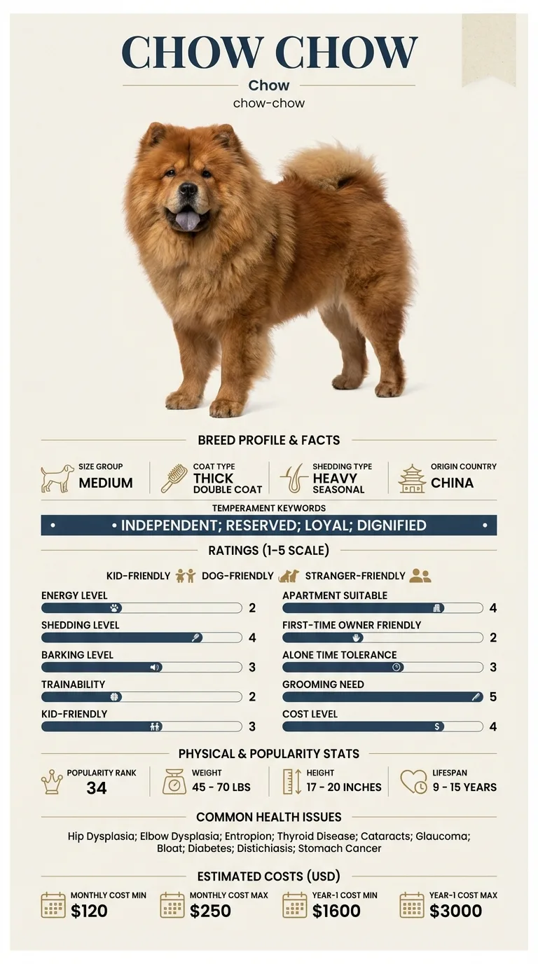
Owners of young Chows, since prolapse most often occurs in the first year or two of life. It is also confusing for owners of a breed that already has documented eyelid disorders, because a red inner corner in a Chow could be several different things.
This guide is not medical advice. If your dog shows pain, sudden behavior change, or worsening symptoms, consult a licensed veterinarian.
Some links may be affiliate links. We may earn a commission at no extra cost to you. We only suggest products we believe are helpful for dog owners.
We research and check what we publish, but we do not warrant that any figure, date, price, or statement is accurate, complete, or current, and we accept no liability for any loss, cost, injury, or other harm arising from the use of this site. Use it as one reference among several. You remain responsible for your own decisions and for your dog's care - confirm anything that matters with a licensed veterinarian or another qualified professional before you act on it. Full disclaimer.
Cherry eye is prolapse of the tear gland of the third eyelid, appearing as a smooth red mass at the inner corner of the eye. It usually occurs in young dogs and does not resolve reliably on its own. It is not among the Chow Chow's documented eye conditions, which are entropion, distichiasis, cataracts and glaucoma, so identification matters before treatment begins.
Common in the breeds where it is documented and uncommon in the Chow Chow, which is not among them. In this breed a red or swollen inner corner is far more likely to reflect an inward-rolling eyelid or abnormal lashes, both of which are documented here. The value of knowing about cherry eye is being able to recognise it if it does occur and knowing which treatment to refuse.
The underlying cause is a congenital weakness in the fibrous attachment holding the gland in place, and the breeds most affected are those in which that attachment is inherently lax. The Chow Chow is not one of them, and its eye conditions run in a different direction entirely - toward lid conformation problems, with entropion and distichiasis both documented. What the Chow's anatomy does contribute is difficulty of examination. Small, deep-set eyes shadowed by a heavy brow and encircled by dense coat mean a lesion at the inner corner can go unnoticed for days.
Environment is not a meaningful cause, but it influences the consequences. A dog that can rub its face freely on carpet or furniture will worsen an exposed gland within hours. Dusty conditions and coat debris carried towards the eye by the surrounding ruff increase irritation. Where a dog is allowed to swim or push through undergrowth with an exposed gland, contamination becomes a genuine risk.
Book within a few days of noticing a prolapsed gland, and sooner if it is enlarging or the dog is rubbing at it. Seek same-day emergency care if the eye is held tightly shut, is visibly cloudy or blue, is bulging, has an obvious ulcer or defect on its surface, or if the dog is in evident pain - these suggest corneal ulceration or raised pressure inside the eye rather than a simple prolapse, and both can destroy vision rapidly. Never instil old or human eye preparations, since some are seriously damaging to an ulcerated cornea.
See all Chow Chow health problems, which breeds are prone to cherry eye, or the full Chow Chow breed guide for temperament, exercise needs and ownership costs.
Surgical repositioning is a day procedure, with a buster collar and medication for around two weeks and a check-up at the end of that. Most glands stay in place, though some swelling persists for a fortnight. Where recurrence happens it is usually within the first few weeks, which is why the post-operative check matters.
Success is a gland back in position, a comfortable eye and normal tear production maintained for life. The last of those is the part that gets overlooked, and it is the reason repositioning rather than removal is the standard. A good outcome is an eye that looks entirely unremarkable and a dog that keeps its own tears.
The Chow Chow's documented eye conditions include entropion, where the lid rolls inward so that hair rubs the eyeball, and distichiasis, where extra lashes grow from the lid margin and abrade the cornea. Both produce redness, discharge and squinting, and both are more common in this breed than gland prolapse is.
What distinguishes cherry eye is the presence of a discrete mass. It is a rounded, smooth, fleshy swelling that sits at the inner corner and partly covers the white of the eye, and it appeared suddenly - often overnight. Generalised redness without a lump points elsewhere.
The breed's anatomy makes this harder to judge than it should be. Deep-set eyes under a heavy brow, framed by a dense ruff, mean an owner may have to physically part the surrounding coat and lift the upper lid to get a proper view. Do it in good light, take a photograph, and do not rely on a glance.
One further note. A Chow with a rolled-in lower lid can have the inner corner tissue pushed forward in a way that superficially resembles a prolapse. Sorting that out is a veterinary job, and the treatments are completely different, which is exactly why guessing is a poor strategy.

This is not usually an out-of-hours emergency, but it is a same-week problem. The gland is exposed tissue and every day it stays out makes it more swollen and harder to replace.
If you think your Chow has cherry eye, the plan is three steps: write down what you have seen and when it started, book a veterinary appointment rather than waiting for the next flare, and take video of the behaviour or symptom before you go — the thing you are worried about rarely happens in the consulting room. Screening in the parents covers hip dysplasia, elbow dysplasia, entropion.
What the vet visit should produce is a diagnosis and a written plan, not just reassurance: what is being ruled out, what the monitoring interval is, and which signs mean you come back sooner. Ask what the treatment costs across a year rather than per visit, because that is the number that decides whether you are managing this condition or reacting to it.
Between appointments, keep a short log — dates, what you saw, what changed. It is the single most useful thing an owner brings to a follow-up, and for cherry eye it is often what separates a clear pattern from a guess.
Bramble was fourteen months old when his owner, Nia, noticed a pink lump at the inner corner of his right eye one Sunday morning. She had almost missed it, because the coat around his eyes was long and the eye itself sat deep under his brow. At the appointment she was told it was a prolapsed third eyelid gland, and that one option was simply to remove it, which would be quicker and cheaper. Nia asked what the consequence would be. The honest answer was a meaningful reduction in tear production and a real chance of dry eye needing lifelong drops. She asked for repositioning instead, and asked whether the left side should be tacked preventively at the same time. Her vet thought that was reasonable given Bramble's age, and both were done under one anaesthetic. Bramble wore a collar for a fortnight, complained about it constantly, and has had no further trouble with either eye in the four years since.
Key takeaway: The gland is worth keeping. If removal is offered as the simple option, ask directly what it costs the dog in tear production, and ask whether the second eye should be secured at the same time so that a breed with a demanding anaesthetic recovery only goes under once.
It is not among the breed's documented conditions, which centre on entropion and distichiasis where the eyes are concerned. Cherry eye is more strongly associated with certain other breeds. That does not make it impossible in a Chow, but a red inner corner in this breed is statistically more likely to be a lid problem.
Occasionally a gland retracts temporarily, which gives owners false reassurance. It almost always comes back, and each episode leaves the gland more swollen. Treat a spontaneous retraction as a reason to still get it examined, not as a resolution.
Because it produces a large share of the eye's tears. Dogs that had the gland removed decades ago frequently developed dry eye, a painful lifelong condition requiring daily medication. The modern approach is to reposition and anchor the gland, preserving its function.
Yes, and in dogs that prolapse one gland the other is at increased risk. Some owners are offered preventive surgery on the unaffected side at the same time, which spares a second anaesthetic. Given that anaesthetic recovery needs careful management in this breed, that argument carries weight.
The gland itself is not richly supplied with pain fibres, but the consequences are uncomfortable - the exposed tissue dries, the eye becomes irritated, and the dog rubs. A dog that is squinting hard, holding the eye shut or in obvious distress has something more than a simple prolapse going on and needs seeing urgently.
Repositioning is usually successful but recurrence does happen and a second procedure is sometimes needed. Ask your vet which technique they use and what their recurrence rate is. A referral to an ophthalmologist is reasonable for a case that has already failed once.
A smooth, rounded pink or red mass at the inner corner of the eye, usually appearing suddenly rather than developing gradually.
No. You can damage the gland and the eye. Stop the dog rubbing, and get it examined within a few days.
It should not be. Removal causes lifelong dry eye. Modern surgery repositions and anchors the gland instead.
Surgical repositioning under general anaesthetic is a mid-range procedure, and doing both eyes together avoids paying twice for anaesthesia.
A one-page prep sheet for this condition: the signs, the questions to ask, what to get priced. Unlocks here.
You get: Vet visit prep sheet
No spam. One-click unsubscribe. See our privacy policy.
Preview page 1 before you decide.