Health
What is the silvery patch on my dog's eye?
If it is in both eyes, symmetrical and painless, corneal dystrophy is likely. A single painful cloudy eye is a different and more urgent problem.

Quick answer
What is the silvery patch on my dog's eye?
If it is in both eyes, symmetrical and painless, corneal dystrophy is likely. A single painful cloudy eye is a different and more urgent problem.
The two questions that sort a cloudy cornea are simple: is it in both eyes, and does it hurt?
Owners who have spotted a silvery oval or a ring of haze in the centre of both of their Berner's eyes and want to know whether it is an emergency, a cataract, or something they can leave alone.
This guide is not medical advice. If your dog shows pain, sudden behavior change, or worsening symptoms, consult a licensed veterinarian.
Some links may be affiliate links. We may earn a commission at no extra cost to you. We only suggest products we believe are helpful for dog owners.
We research and check what we publish, but we do not warrant that any figure, date, price, or statement is accurate, complete, or current, and we accept no liability for any loss, cost, injury, or other harm arising from the use of this site. Use it as one reference among several. You remain responsible for your own decisions and for your dog's care - confirm anything that matters with a licensed veterinarian or another qualified professional before you act on it. Full disclaimer.
Corneal dystrophy is an inherited accumulation of material within the layers of the clear front window of the eye. It typically appears in both eyes in a similar position and pattern, produces no pain and no discharge, and often has no effect on how the dog sees or behaves.
Not documented as a breed condition in the Bernese Mountain Dog, whose recognised eye issue is progressive retinal atrophy. Corneal deposits are found across many breeds and are one of the more frequent incidental findings at routine eye examinations.
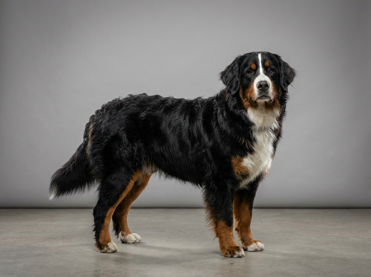
There is no specific Bernese predisposition here, and it should not be confused with the retinal disease that is on the breed's documented list. The one genuinely breed-relevant point is that a Bernese's expressive rust-coloured facial markings and the hair around the eyes can obscure the eye surface, so deposits are often noticed later than in a short-coated, light-faced dog.
Environment plays little part in causing deposits, but it plays a large part in the complications worth watching for. A breed that walks in long grass, works outdoors and pushes its face into hedgerows collects scratches, and a corneal surface with an existing deposit is one that merits prompt attention when it is injured.
Go the same day for an eye that is held partly closed, watering heavily, red, or being rubbed, and for cloudiness that has appeared in one eye only or over the space of a day or two. Those signs point to a corneal ulcer or to inflammation inside the eye, and an ulcer in a large outdoor dog can deepen fast enough to threaten the eye. Book a routine appointment for symmetrical, painless haze so it can be identified, and mention any history of raised cholesterol, thyroid problems or previous eye inflammation, since those can be relevant to the type of deposit present.
There is little to do about corneal dystrophy itself, which makes it easy to do the wrong thing out of a wish to act.
Start here: Bernese Mountain Dog Most Common Genetic Disorders
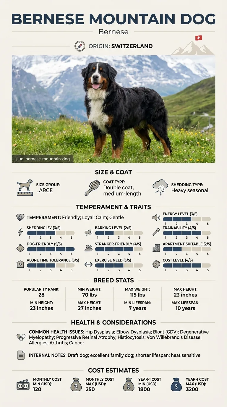
See all Bernese Mountain DOG health problems, which breeds are prone to corneal dystrophy, or the full Bernese Mountain DOG breed guide for temperament, exercise needs and ownership costs.
Deposits typically change very slowly, over years rather than months, and many remain static for a dog's whole life. Monitoring is usually annual, folded into a routine health examination.
A correctly identified, painless finding that needs no treatment, an owner who can distinguish it from an injury, and an eye that stays comfortable.
Deposit is the first. Material accumulates in the corneal layers, most often appearing as a symmetrical oval, ring or crystalline patch toward the centre of both eyes. It is typically silvery or grey-white, sharply outlined, and the dog behaves entirely normally. Vision is rarely affected because the deposit does not usually cover enough of the visual axis to matter. This is corneal dystrophy, and in most dogs it needs no treatment at all, only recording so that change over years can be tracked.
Swelling is the second. When the cell layer lining the inside of the cornea stops pumping fluid out, the tissue takes up water and becomes diffusely blue and hazy rather than sharply outlined. This looks quite different: cloudier, softer-edged and more like frosted glass. It can progress and can eventually cause discomfort, so it needs veterinary follow-up rather than observation alone.
Injury is the third and the one that matters most acutely. A scratch or ulcer on the corneal surface produces a painful eye: held partly shut, watering, red, and rubbed against furniture. There may be a focal grey or white spot rather than a symmetrical pattern. This is urgent, because a corneal ulcer in a large dog on a farm walk can deepen quickly.
The home test is therefore behavioural rather than visual. Watch whether your dog is squinting, whether both eyes look the same, and whether there is any discharge. A relaxed, comfortable dog with matched silvery patches in both eyes is a very different situation from a dog with one weeping eye it will not open.

Wilhelm noticed a silvery oval in his Berner Trudi's right eye during a photograph, then found a near-identical one in the left. He booked an appointment expecting bad news. His vet examined both eyes, applied a stain to confirm the surfaces were intact, and identified symmetrical corneal deposits with no ulceration and no pain. Trudi needed nothing. Wilhelm was shown how to check for squinting and watering, and asked to photograph the eyes twice a year. Two years on, the ovals are the same size and Trudi has never had a problem with them.
Key takeaway: Not every visible change in an eye is a developing disease. The examination that confirmed nothing was needed was still worth making, because it told Wilhelm exactly what would change the situation.
Usually very little. The deposits typically occupy a small part of the cornea and light passes around them. Where an unusually extensive deposit does interfere, a veterinary ophthalmologist can assess whether anything is worth doing, but intervention is uncommon.
Position. A cataract is an opacity in the lens, which sits behind the pupil, so it appears within the dark circle of the eye. Corneal changes are on the front surface and can appear anywhere across it, including over the coloured iris. A vet can distinguish them instantly.
No. Uncomplicated corneal dystrophy causes no discomfort, which is one of its defining features. A dog squinting, watering or rubbing has something else going on, most likely a corneal injury or an inflammatory condition, and that needs prompt attention.
There is no treatment that removes the deposits reliably, and most dogs need none. Where the surface breaks down over a deposit, which is uncommon, treatment addresses that complication. Your vet may recommend periodic examination rather than any active intervention.
Sometimes. Fatty corneal deposits can occur alongside disorders of fat metabolism or thyroid function, so your vet may suggest bloodwork, particularly if the deposits appeared suddenly or the dog has other signs. That is a screening question rather than an alarming one.
It is not among the breed's documented health issues, which include progressive retinal atrophy as the recognised eye condition. Corneal dystrophy occurs across many breeds, and any dog with corneal cloudiness should be examined rather than assessed against a breed list.
If it is in both eyes, symmetrical and painless, corneal dystrophy is likely. A single painful cloudy eye is a different and more urgent problem.
Squinting, watering, redness, rubbing, or cloudiness in only one eye. Corneal injury can deepen quickly and needs same-day attention.
Not without veterinary direction. Some preparations are harmful when the corneal surface is damaged, and you cannot assess that at home.
Yes, lightly. Photograph both eyes a few times a year in the same light so any change in size or character is documented.
Usually very little. It is typically a diagnosis made at a routine examination, needing observation rather than treatment.
A one-page prep sheet for this condition: the signs, the questions to ask, what to get priced. Unlocks here.
You get: Vet visit prep sheet
No spam. One-click unsubscribe. See our privacy policy.
Preview page 1 before you decide.