Health
What is the pink lump in my dog's inner eye corner?
Almost certainly a prolapsed third eyelid tear gland. It is not a tumour, but it needs veterinary assessment within a day or two.

Quick answer
What is the pink lump in my dog's inner eye corner?
Almost certainly a prolapsed third eyelid tear gland. It is not a tumour, but it needs veterinary assessment within a day or two.
The pink lump is a working tear gland in the wrong place. Where it goes next determines whether the eye stays comfortable for life.
Owners of young Berners, usually under two, who have found a red swelling at the inner corner of one eye that appeared over a day and are trying to work out how urgent it is.
This guide is not medical advice. If your dog shows pain, sudden behavior change, or worsening symptoms, consult a licensed veterinarian.
Some links may be affiliate links. We may earn a commission at no extra cost to you. We only suggest products we believe are helpful for dog owners.
We research and check what we publish, but we do not warrant that any figure, date, price, or statement is accurate, complete, or current, and we accept no liability for any loss, cost, injury, or other harm arising from the use of this site. Use it as one reference among several. You remain responsible for your own decisions and for your dog's care - confirm anything that matters with a licensed veterinarian or another qualified professional before you act on it. Full disclaimer.
A small band of tissue normally anchors the third eyelid's tear gland in position at the inner corner of the eye. When that anchoring is weak, the gland flips forward and sits on the surface as a pink or red swelling. It keeps producing tears but is exposed, dries out and becomes inflamed.
Not a documented Bernese Mountain Dog condition, and far more strongly associated with certain other breeds. It is nonetheless something any dog can develop, and it is one of the more recognisable eye problems an owner will ever see, which is why it generates immediate concern.
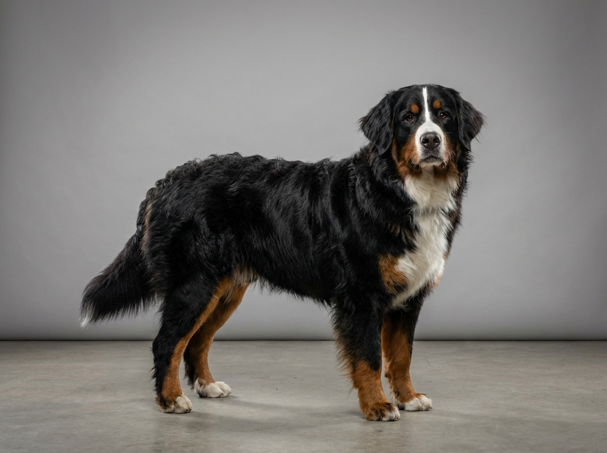
This is a connective tissue problem rather than a size or coat problem, and the Bernese is not among the breeds where it is common. Where it does occur in a Berner, the relevant breed factors are practical rather than anatomical: a large young dog is harder to keep from rubbing its face, and the eye area is partly obscured by the rust-coloured facial markings until you look closely.
Anything that makes a dog rub its face increases the chance of a weakly anchored gland slipping forward. Allergic irritation, dust, wind on country walks and a habit of pushing the face into undergrowth all contribute. A young dog in a household with rough play is more likely to take a knock to the face.
Book within one to two days for a newly visible pink swelling at the inner corner of the eye. Go the same day if the eye is being held closed, if there is a green or yellow discharge, if the surface of the eye itself looks cloudy or dull, or if your dog is rubbing so persistently that you cannot prevent it, because corneal damage happens quickly and is genuinely painful. A first-time prolapse in a dog over four years old also deserves prompter attention, since a mass behind the third eyelid has to be excluded rather than assumed away.
This is a condition where a well-meaning owner can do real harm in the first forty-eight hours.
Start here: Bernese Mountain Dog Most Common Genetic Disorders
See all Bernese Mountain DOG health problems, which breeds are prone to cherry eye, or the full Bernese Mountain DOG breed guide for temperament, exercise needs and ownership costs.
Surgery is usually scheduled within days to a couple of weeks. Healing takes one to two weeks with a cone, and the eye looks normal shortly afterwards. A minority recur and need a second procedure.
A gland sitting where it belongs, normal tear production preserved, and a comfortable eye that needs no ongoing medication.
The third eyelid gland is not a spare part. It produces a large proportion of the watery component of the tear film, and the tear film is what keeps the cornea clear, lubricated and protected from infection. Remove the gland and you leave a young dog with permanently reduced tear production, which frequently develops into dry eye: a chronically sore, red, sticky eye needing lifelong medication several times daily and carrying a risk of ulceration and scarring.
Modern surgical repair therefore aims to reposition the gland and secure it in place, either by creating a pocket in the third eyelid to tuck it into or by anchoring it back to the surrounding tissue. These procedures are routine for a veterinary ophthalmologist and are widely performed in general practice too. A small proportion recur and need a second attempt, which is a better problem than a lifetime of dry eye.
Before surgery, the aim at home is to keep the exposed tissue moist and untouched. Your vet may prescribe lubricant drops and an anti-inflammatory preparation, and a cone stops the dog rubbing. This is a matter of days rather than weeks; the longer the gland sits exposed, the more it swells and dries.
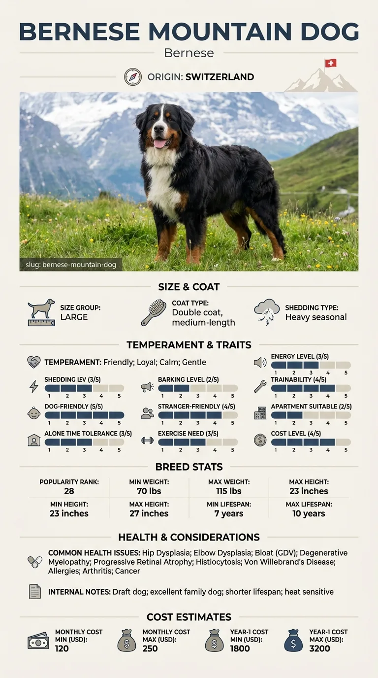
The condition is most associated with certain young breeds and is not among the Bernese Mountain Dog's documented health issues, which centre on hips and elbows, bloat, degenerative myelopathy, progressive retinal atrophy, histiocytosis, bleeding disorder, allergies, arthritis and cancer. It occurs in this breed as it can in almost any dog, and where it does the management is identical.

If you think your Bernese has cherry eye, the plan is three steps: write down what you have seen and when it started, book a veterinary appointment rather than waiting for the next flare, and take video of the behaviour or symptom before you go — the thing you are worried about rarely happens in the consulting room. Screening in the parents covers hip dysplasia, elbow dysplasia, bloat (gdv).
What the vet visit should produce is a diagnosis and a written plan, not just reassurance: what is being ruled out, what the monitoring interval is, and which signs mean you come back sooner. Ask what the treatment costs across a year rather than per visit, because that is the number that decides whether you are managing this condition or reacting to it.
Between appointments, keep a short log — dates, what you saw, what changed. It is the single most useful thing an owner brings to a follow-up, and for cherry eye it is often what separates a clear pattern from a guess.
Bex was fourteen months old when her owner Callum found a red bulge at the inner corner of her right eye on a Saturday morning. He photographed it, phoned on Monday, and was offered a slot that week. The conversation he almost did not have was about technique. He asked whether the gland would be removed or repositioned, and his vet explained the pocket technique and why removal is avoided. Bex had the gland tucked back and secured, wore a cone for twelve days, and has had no further trouble. Her left gland prolapsed nine months later and was repaired the same way.
Key takeaway: One question at the booking stage was the whole difference between a fortnight in a cone and a lifetime of drops for dry eye.
Initially it is more irritating than painful, but exposed glandular tissue dries out and becomes inflamed, and dogs then rub at it, which causes real discomfort and can damage the cornea. It is not an emergency in the way an acutely painful eye is, but it should be seen within a day or two.
Occasionally a gland slips back temporarily, which gives false reassurance, but permanent spontaneous resolution is uncommon. Prolapse tends to recur and to become more swollen with each episode, so surgical repositioning is the standard recommendation.
Because it produces a large share of the eye's tears. Removal is associated with dry eye later in life, which means daily medication indefinitely and a risk of corneal ulceration. Repositioning preserves function, and it is what most vets and all ophthalmologists now recommend.
Most cases appear in dogs under two years old, and often before one. A first-time prolapse in a middle-aged or older dog is less typical and warrants a careful look for another cause, such as a mass or inflammation behind the third eyelid.
Yes. A dog that has prolapsed one gland has a raised chance of the other following, sometimes months later. Keep an eye on the unaffected side, and mention the history if it happens so your vet knows this is a second event rather than a first.
Costs vary by region and by whether it is done in general practice or by a specialist, and both eyes together cost more than one. Recovery is straightforward: a cone for one to two weeks, prescribed drops, and no swimming or rough play while it heals.
Almost certainly a prolapsed third eyelid tear gland. It is not a tumour, but it needs veterinary assessment within a day or two.
No. You risk damaging the gland and introducing infection, and it will not stay. Keep the dog from rubbing and book an appointment.
Repositioning. Removing the gland is associated with lifelong dry eye. Ask your vet directly which technique they use.
A well-fitted cone, and remove access to carpets and furniture the dog likes to rub its face against. Supervise closely until surgery.
It is a modest surgical cost compared with treating lifelong dry eye, which needs daily medication for years. Fixing it properly is the cheaper path.
A one-page prep sheet for this condition: the signs, the questions to ask, what to get priced. Unlocks here.
You get: Vet visit prep sheet
No spam. One-click unsubscribe. See our privacy policy.
Preview page 1 before you decide.