Health
Is corneal dystrophy the same as cataracts?
No, corneal dystrophy affects the cornea's surface layer, while cataracts cloud the lens deeper in the eye.

Quick answer
Is corneal dystrophy the same as cataracts?
No, corneal dystrophy affects the cornea's surface layer, while cataracts cloud the lens deeper in the eye.
A hazy patch on the surface of your dog's eye that doesn't seem to be affecting their vision is more likely corneal dystrophy than the more commonly discussed cataracts.
This is most relevant to owners who've noticed a cloudy patch and are trying to figure out whether it's the more serious, vision-threatening cataracts documented in this breed, or the generally more benign corneal dystrophy.
This guide is not medical advice. If your dog shows pain, sudden behavior change, or worsening symptoms, consult a licensed veterinarian.
Some links may be affiliate links. We may earn a commission at no extra cost to you. We only suggest products we believe are helpful for dog owners.
We research and check what we publish, but we do not warrant that any figure, date, price, or statement is accurate, complete, or current, and we accept no liability for any loss, cost, injury, or other harm arising from the use of this site. Use it as one reference among several. You remain responsible for your own decisions and for your dog's care - confirm anything that matters with a licensed veterinarian or another qualified professional before you act on it. Full disclaimer.
Check this first
Corneal dystrophy itself is rarely an emergency and often requires monitoring rather than active treatment. However, sudden eye pain, squinting, excessive tearing, redness, or a rapidly worsening cloudy patch can indicate a corneal ulcer or erosion, which is a genuinely urgent, painful condition requiring same-day veterinary attention.
The practical difference to watch for is speed and discomfort: corneal dystrophy typically develops slowly and doesn't cause pain, while an ulcer develops faster and is uncomfortable. If your dog is suddenly squinting, pawing at an eye, or clearly uncomfortable, treat it as urgent regardless of what the underlying cause turns out to be.
If any of these apply, call your vet or an emergency clinic now. The rest of this page is for the cases that are not an emergency.
Corneal dystrophy involves deposits building up in the cornea, the eye's clear outer surface, distinct from the lens-clouding cataracts that are a documented issue in this breed. It's generally a slower-progressing, less vision-threatening condition, often managed with monitoring rather than active treatment.
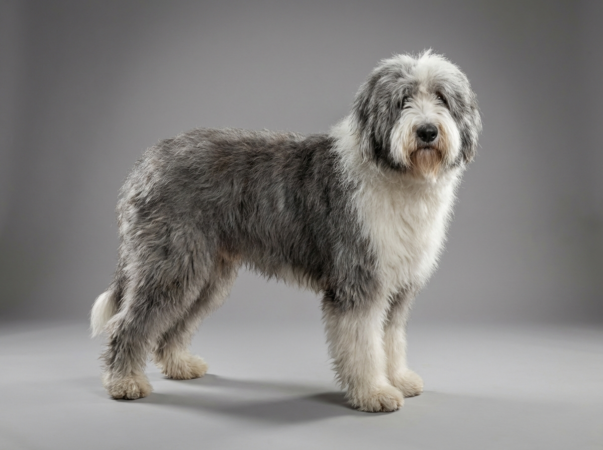
Corneal dystrophy is recognized across many dog breeds and isn't specifically documented as elevated in Old English Sheepdogs. It's included here mainly because it's frequently confused with this breed's documented cataract risk.
There's no strong documented breed-specific link between Old English Sheepdogs and corneal dystrophy. This breed's documented eye risk is cataracts, a lens condition, which is structurally and clinically distinct from corneal dystrophy despite both being described loosely as eye cloudiness.
Environmental factors play a limited role in true corneal dystrophy, which is more commonly linked to genetics or, in some cases, underlying metabolic factors, rather than external triggers.
Talk to your vet about any new cloudiness noticed in the eye to confirm what you're dealing with. Seek same-day care for sudden pain, squinting, redness, or excessive tearing, which points toward a different, more urgent issue.
Since the two conditions look superficially similar but behave very differently, mixing them up leads to unnecessary worry or, less often, missed concern.
See all OLD English Sheepdog health problems, which breeds are prone to corneal dystrophy, or the full OLD English Sheepdog breed guide for temperament, exercise needs and ownership costs.
Corneal dystrophy is often stable for long periods and monitored with periodic eye exams rather than treated actively. Any needed diagnostic workup to distinguish it from cataracts is usually resolved within one or two vet visits.
Success looks like a clear diagnosis distinguishing this from cataracts, and confidence that a cosmetic finding doesn't require the more involved management a lens condition would.
Rosie's owner noticed a small hazy patch on the surface of one eye and, having read about this breed's documented cataract risk, worried it meant surgery was in her future. A vet exam with specialized lighting showed the cloudiness sat within the cornea itself, not the lens, consistent with corneal dystrophy rather than a cataract. Rosie's vet recommended monitoring with periodic checks rather than any active treatment, and the patch has remained stable for over a year since.
Key takeaway: Not every cloudy-looking eye finding is a cataract. A proper exam distinguishing cornea from lens changes the whole picture, and often the level of concern warranted.
Corneal dystrophy involves deposits building up in the cornea, the clear surface layer at the front of the eye, while cataracts cloud the lens deeper inside the eye. Corneal dystrophy is often more cosmetic and slower to affect vision; cataracts, a documented issue in this breed, can more significantly impair vision as they progress.
Typically not. It's usually a slow, non-painful change. Sudden pain, squinting, or redness points toward a different, more urgent issue like a corneal ulcer, rather than dystrophy itself.
Many cases are simply monitored over time rather than actively treated, since they don't typically progress to significantly impair vision. Your vet will guide whether monitoring or another approach is appropriate for your specific dog.
Through a close eye exam, often with specialized lighting, that lets a vet or veterinary ophthalmologist see exactly where the cloudiness sits, in the cornea versus the lens, which is the key distinguishing factor from cataracts.
It can in some cases, particularly if deposits are extensive or centrally located, but it's generally less likely to significantly impair vision than cataracts, which affect the lens directly.
If there's any uncertainty about which condition you're dealing with, a referral to a veterinary ophthalmologist for a more detailed exam is a reasonable step, especially given the different implications of each condition.
Some corneal conditions in dogs are believed to have a hereditary component, though this isn't a well-documented specific predisposition in this breed the way cataracts are.
No, corneal dystrophy affects the cornea's surface layer, while cataracts cloud the lens deeper in the eye.
Often it's a cosmetic, slowly progressing finding that doesn't significantly threaten vision, unlike cataracts.
Typically not; sudden pain or squinting points toward a different, more urgent issue like a corneal ulcer.
With a close eye exam, sometimes by a veterinary ophthalmologist, to confirm the cloudiness is in the cornea rather than the lens.
Yes, periodic monitoring is reasonable to confirm it stays stable rather than progressing.
A one-page prep sheet for this condition: the signs, the questions to ask, what to get priced. Unlocks here.
You get: Vet visit prep sheet
No spam. One-click unsubscribe. See our privacy policy.
Preview page 1 before you decide.