Health
What is the pink lump in my dog's inner eye corner?
Almost certainly the prolapsed tear gland of the third eyelid, known as cherry eye. It needs veterinary confirmation and surgical replacement.

Quick answer
What is the pink lump in my dog's inner eye corner?
Almost certainly the prolapsed tear gland of the third eyelid, known as cherry eye. It needs veterinary confirmation and surgical replacement.
A red bead has appeared in the corner of your dog's eye overnight. It is not a tumour, it is not an injury, and the wrong treatment is the one that makes it disappear fastest.
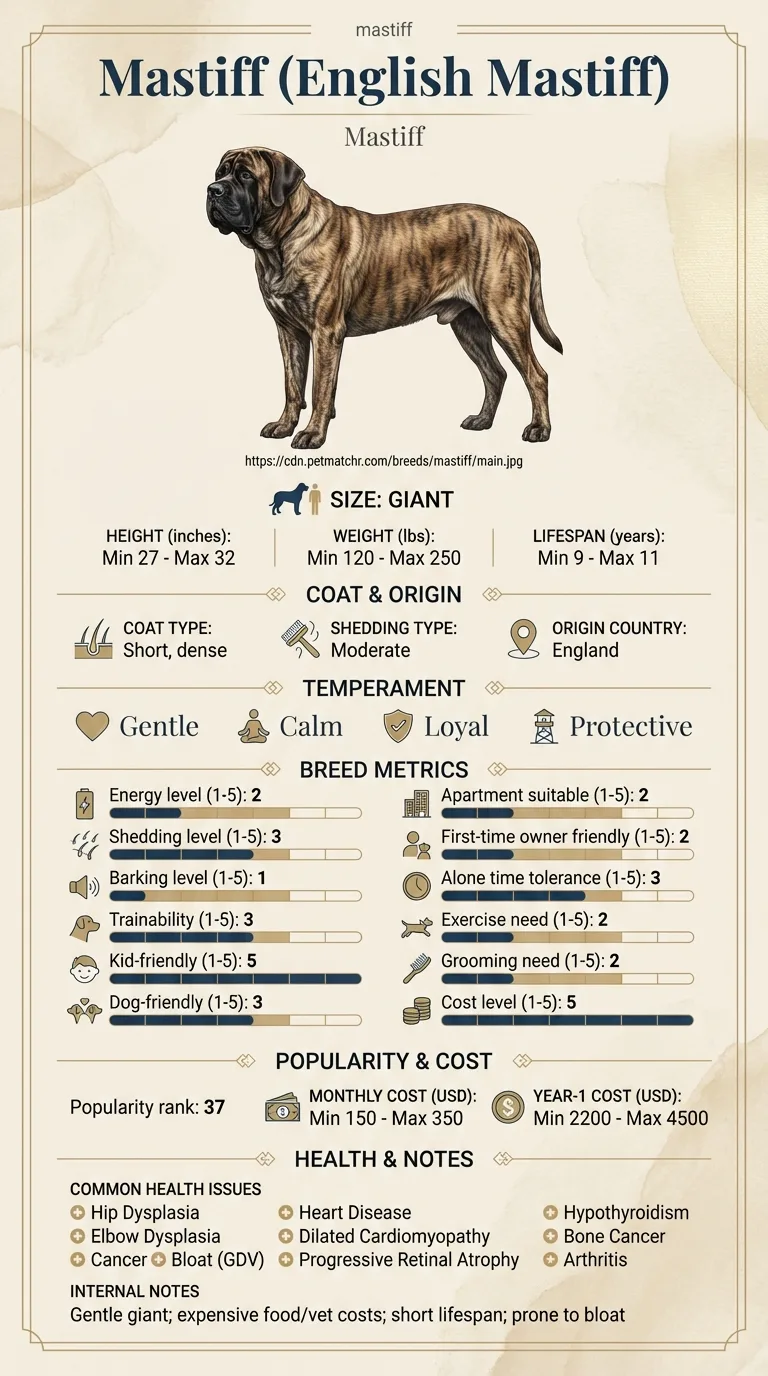
Owners of young Mastiffs, usually under two, who have woken up to a pink lump that was not there yesterday, and owners weighing a vet's recommendation to tack the gland back against an older or cheaper suggestion to simply cut it out.
This guide is not medical advice. If your dog shows pain, sudden behavior change, or worsening symptoms, consult a licensed veterinarian.
Some links may be affiliate links. We may earn a commission at no extra cost to you. We only suggest products we believe are helpful for dog owners.
We research and check what we publish, but we do not warrant that any figure, date, price, or statement is accurate, complete, or current, and we accept no liability for any loss, cost, injury, or other harm arising from the use of this site. Use it as one reference among several. You remain responsible for your own decisions and for your dog's care - confirm anything that matters with a licensed veterinarian or another qualified professional before you act on it. Full disclaimer.
Cherry eye is prolapse of the tear-producing gland that normally sits hidden behind the third eyelid. The connective tissue anchoring it gives way and the gland flips forward, appearing as a smooth pink or red swelling in the inner corner of the eye. The gland is not diseased. It is simply in the wrong place, and left there it dries, swells and stops working properly.
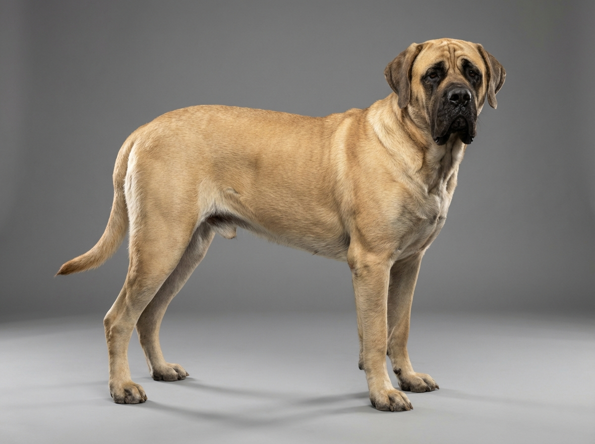
Cherry eye is common across dogs as a whole but is strongly concentrated in particular breeds, and the English Mastiff is not among the classic ones. It is not on the breed's documented health issue list. It appears here because loose facial tissue makes it possible in any Mastiff, and because the wrong first decision, removal instead of replacement, has consequences that last the dog's whole life.
The mechanism is a failure of the small fibrous attachment that holds the gland behind the third eyelid. Breeds with generous, loose facial skin and less tightly fitting eyelids are over-represented, and the English Mastiff has a heavy head with a degree of natural lid looseness, which makes it structurally possible even though the breed is not a classic candidate. Because the anchoring tissue is a developmental feature, cases cluster in young dogs rather than appearing with age.
There is little environmental contribution to whether the gland prolapses in the first place. What the environment does influence is what happens afterwards. A dog with access to rough carpet, hedges and its own paws will rub an irritated eye and cause corneal damage that would not otherwise occur. Dusty or smoky rooms inflame the exposed tissue further. Prompt use of a protective collar, unglamorous as it is, prevents most secondary damage.
Book an appointment as soon as you notice the swelling, ideally within a few days, because an exposed gland deteriorates and becomes harder to replace. Seek same-day attention if the eye becomes red and painful, if the dog is squinting or holding it shut, if there is thick or coloured discharge, if the surface of the eye looks cloudy or dull, or if the dog is rubbing persistently despite a collar. A corneal ulcer developing under a prolapsed gland is a genuine urgency, because ulcers can deepen quickly. Never attempt to push the gland back into place yourself.
See all Mastiff health problems, which breeds are prone to cherry eye, or the full Mastiff breed guide for temperament, exercise needs and ownership costs.
Surgery is usually performed within one to three weeks of diagnosis. Swelling settles over about a fortnight, with the protective collar worn for one to two weeks and drops continuing a little longer. Most dogs look entirely normal within a month. If the gland recurs it typically does so within the first few weeks, and the second eye, if it goes, usually does so within a year.
Success is a gland sitting back where it belongs, a comfortable eye with no discharge or squinting, an intact cornea, and normal tear production in later life. The measure of success here is largely invisible: a dog in its senior years that does not need ointment twice a day is the return on a decision made when it was eighteen months old.
The instinct on seeing a lump is to have it taken away. In this specific case that instinct trades a cosmetic problem now for a serious one later.
Cherry eye typically appears suddenly, often in a young dog, and frequently in one eye first with the other following within months. Owners describe finding it fully formed on waking.
The first step is examination, not surgery. Your vet needs to confirm that the swelling really is the prolapsed gland and not a mass, a foreign body, or inflamed conjunctiva, and to check that the cornea has not been damaged by the exposed tissue rubbing against it. In a breed with loose facial skin and a degree of eyelid laxity, the surrounding lid conformation is worth assessing at the same time, since a rolled or drooping lid can complicate both the problem and the repair.
Surgery is usually scheduled promptly rather than urgently. The delay matters because an exposed gland dries out, becomes inflamed and swollen, and a swollen gland is harder to replace neatly. Some vets prescribe drops or ointment in the interval to keep the tissue moist and reduce inflammation, which makes the operation easier.
The procedure itself creates a pocket in the third eyelid tissue and buries the gland inside it, or anchors it back to the deeper tissue of the socket. It is done under general anaesthesia, which in a giant breed is worth planning properly, and it is usually a day procedure. Aftercare means a protective collar for a week or two, drops, and preventing a large dog from rubbing its face on the carpet, which is harder than it sounds.
One honest caution: recurrence rates are not zero, and the second eye prolapsing within a year is common enough that many owners end up going through this twice. Neither of those outcomes changes the underlying advice, which is that the gland belongs inside the eye.

If you think your Mastiff has cherry eye, the plan is three steps: write down what you have seen and when it started, book a veterinary appointment rather than waiting for the next flare, and take video of the behaviour or symptom before you go — the thing you are worried about rarely happens in the consulting room. Screening in the parents covers hip dysplasia, elbow dysplasia, cancer.
What the vet visit should produce is a diagnosis and a written plan, not just reassurance: what is being ruled out, what the monitoring interval is, and which signs mean you come back sooner. Ask what the treatment costs across a year rather than per visit, because that is the number that decides whether you are managing this condition or reacting to it.
Between appointments, keep a short log — dates, what you saw, what changed. It is the single most useful thing an owner brings to a follow-up, and for cherry eye it is often what separates a clear pattern from a guess.
Nadia's Mastiff Rufus developed a pink swelling in his left inner eye corner at fourteen months. The first opinion she sought suggested simply removing the gland, which was quicker, cheaper and would resolve the appearance immediately. Something about it did not sit right, and she asked for a second opinion. The second vet explained what the gland actually did, and that around a third of the eye's tear production sat inside the lump Nadia was being offered a discount to remove. Rufus had a pocketing procedure instead. The gland stayed put. Eight months later the right eye prolapsed and was repaired the same way, which Nadia had been warned to expect and had budgeted for. Rufus is seven now and has never needed eye medication. Nadia's summary is that the expensive option was the cheap one, because the alternative was ointment twice a day for a decade.
Key takeaway: Cherry eye is one of the few situations where the faster, cheaper surgical option is clearly the worse one. The gland is not a lump to be removed, it is a working part of the eye.
Initially it is more of an irritation than a pain, and many dogs behave entirely normally at first. It becomes painful once the exposed gland dries and swells, or once it rubs the cornea enough to cause an ulcer. A dog that starts squinting, rubbing or producing discharge has moved past the comfortable stage.
Occasionally a gland pops back temporarily, particularly early on, and owners understandably take that as resolution. It almost always reappears. A gland that has prolapsed once has lost its normal attachment, and the fix is surgical rather than spontaneous.
Do not try. You risk damaging both the gland and the cornea, and unclean fingers around an exposed mucous surface invite infection. Warm moist compresses and vet-prescribed lubrication while you wait for the appointment are appropriate. Physical manipulation is not.
Dry eye, or keratoconjunctivitis sicca, is inadequate tear production. The surface of the eye becomes inflamed and painful, thick discharge forms, ulcers recur, and pigment gradually spreads across the cornea reducing vision. It is treatable but not curable, and it demands medication several times a day for the rest of the dog's life.
Frequently, yes. Many dogs that prolapse one gland prolapse the other within a year. It is worth budgeting for that possibility and worth asking your vet whether they would recommend addressing the second side preventively, which is a decision with arguments on both sides.
Cherry eye is most strongly associated with Bulldogs, Cocker Spaniels, Beagles and similar breeds, and it is not among the documented health issues for the English Mastiff, whose recognised eye concern is progressive retinal atrophy. Any breed with loose facial and eyelid tissue can produce it, which is why it is worth recognising.
It is a relatively modest procedure by veterinary surgical standards, but in a giant breed the anaesthetic component pushes the total up, and doing both eyes doubles it. Compare that once-off cost against a lifetime of twice-daily prescription ointment for dry eye, which is what removal typically leads to.
Almost certainly the prolapsed tear gland of the third eyelid, known as cherry eye. It needs veterinary confirmation and surgical replacement.
No. Removal substantially raises the risk of lifelong dry eye. The current standard of care is to replace and anchor the gland.
Prompt rather than emergency, unless the eye becomes painful, red or the dog is squinting, in which case seek same-day attention.
Only with lubrication your vet has prescribed and a collar to stop rubbing. Never push the gland back manually.
Often, within about a year. Budget for the possibility and discuss it with your vet at the first surgery.
A one-page prep sheet for this condition: the signs, the questions to ask, what to get priced. Unlocks here.
You get: Vet visit prep sheet
No spam. One-click unsubscribe. See our privacy policy.
Preview page 1 before you decide.