Health
What does glaucoma look like in a dog's eye?
A red, sometimes cloudy or blue-tinged eye with an enlarged pupil and visible discomfort, such as squinting or rubbing at the face.

Quick answer
What does glaucoma look like in a dog's eye?
A red, sometimes cloudy or blue-tinged eye with an enlarged pupil and visible discomfort, such as squinting or rubbing at the face.
A red, painful eye in a dog is never just irritation until pressure has been ruled out. By the time vision is visibly affected, damage may already be permanent.
Owners whose dog suddenly develops a red, cloudy or painful-looking eye, and anyone managing a dog already diagnosed with glaucoma in one eye and trying to protect the other.
This guide is not medical advice. If your dog shows pain, sudden behavior change, or worsening symptoms, consult a licensed veterinarian.
Some links may be affiliate links. We may earn a commission at no extra cost to you. We only suggest products we believe are helpful for dog owners.
We research and check what we publish, but we do not warrant that any figure, date, price, or statement is accurate, complete, or current, and we accept no liability for any loss, cost, injury, or other harm arising from the use of this site. Use it as one reference among several. You remain responsible for your own decisions and for your dog's care - confirm anything that matters with a licensed veterinarian or another qualified professional before you act on it. Full disclaimer.
Glaucoma is a rise in pressure inside the eye caused by impaired drainage of the fluid the eye continuously produces. Sustained pressure damages the optic nerve within hours, causing pain and permanent vision loss if not treated urgently. It is the most time-critical eye condition affecting this breed, distinct from the slower, often painless conditions elsewhere on the documented list.
Glaucoma occurs across dog breeds generally and is not confined to any single breed profile. It is included here because raised eye pressure is one of the unrelated conditions grouped under this breed's broader ocular disease picture, and because its urgency differs so sharply from the breed's more commonly discussed eye conditions that it deserves separate, dedicated attention.
There is no documented breed-specific structural predisposition driving glaucoma in the Bernese Mountain Dog the way lid conformation or inherited retinal degeneration are documented elsewhere. What matters for this breed is context: because eye pressure emergencies can be mistaken for the breed's more familiar, slower eye conditions, recognising the difference in urgency is the main breed-relevant point.
Any prior eye injury, inflammation, or other eye disease affecting the drainage angle can set the stage for secondary glaucoma later. Access to prompt veterinary or emergency care, given how quickly pressure damage becomes permanent, is the most significant environmental factor in the actual outcome.
Treat any sudden redness, cloudiness, visible pain, or pupil change in one eye as a same-day emergency, not a routine appointment. This is one of the few conditions on this breed's profile where hours genuinely matter, because pressure-related optic nerve damage becomes permanent quickly and cannot be reversed once it has occurred.
Because glaucoma can look like several less urgent conditions in its early hours, most of the damage done by delay comes from reasonable-seeming decisions to wait and watch.
Start here: Bernese Mountain Dog Most Common Genetic Disorders
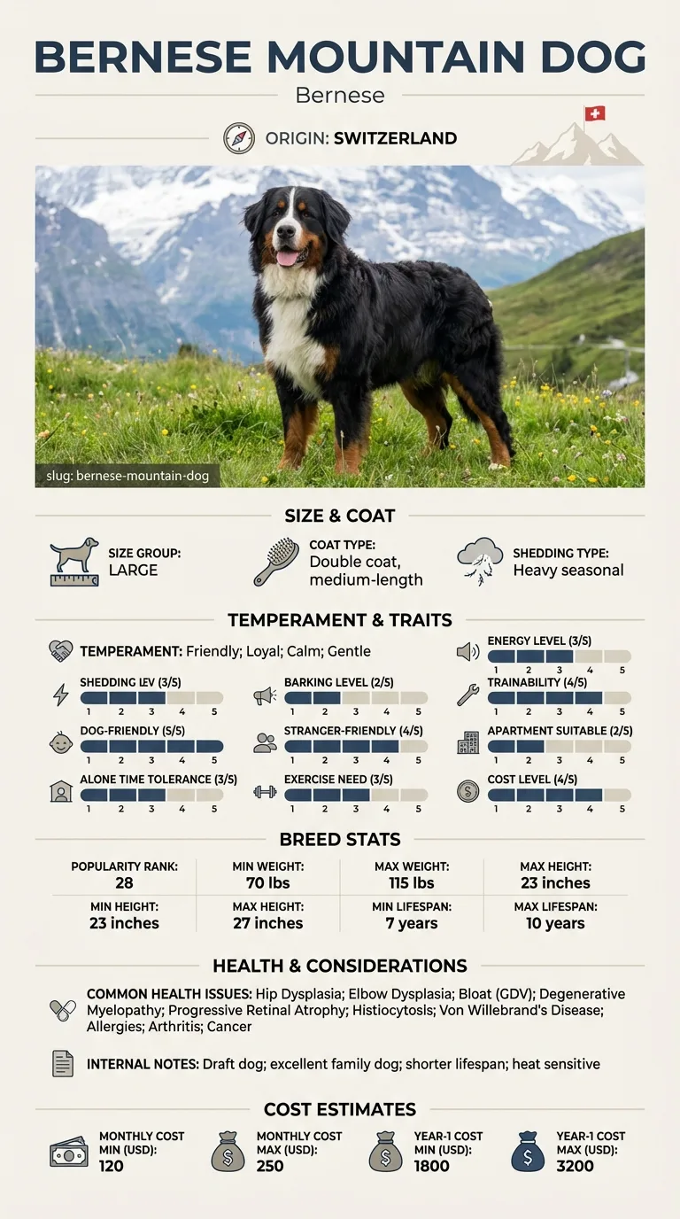
See all Bernese Mountain DOG health problems, which breeds are prone to glaucoma, or the full Bernese Mountain DOG breed guide for temperament, exercise needs and ownership costs.
An acute pressure spike needs treatment within hours to protect vision. Once stabilised, chronic glaucoma typically requires lifelong monitoring and daily medication rather than a defined recovery period.
Success in an acute episode is preserved vision and a controlled pressure reading on recheck. For chronic cases, success is stable pressure maintained through consistent medication rather than a cure, and vigilant monitoring of the second eye if only one has been affected so far.
The eye continuously produces a clear fluid that nourishes internal structures and drains away through a specific angle at the front of the eye. Glaucoma occurs when that drainage angle becomes blocked or impaired, so fluid keeps being produced but can no longer leave at the same rate. Pressure inside the eye rises, and because the eye is a closed, fluid-filled sphere, that rising pressure is transmitted directly onto the optic nerve and retina at the back of the eye.
The optic nerve is not tolerant of sustained pressure. Nerve fibres begin to die within hours of a severe pressure spike, and once dead they do not regenerate, which is what makes this fundamentally different from every other eye condition documented for this breed. A cataract can be monitored and eventually operated on without the same time pressure. Corneal dystrophy causes no functional loss at all. Glaucoma is a race against permanent, irreversible nerve damage from the moment pressure rises significantly.
What an owner actually sees varies. A classic presentation includes a red eye, a cloudy or blueish cornea from fluid pushed into the tissue, an enlarged-looking pupil that responds poorly to light, and visible pain: squinting, rubbing at the face, reluctance to have the head touched, or a general drop in activity and appetite. Some cases develop more gradually with subtler signs, which is part of why any change in one eye compared with the other deserves prompt professional assessment rather than a wait-and-see approach.
Diagnosis is made with a tonometer, a simple instrument that measures pressure directly at the vet's office in seconds. Treatment focuses on lowering pressure urgently with medication, and in some cases surgery, to protect remaining vision, while chronic long-term glaucoma is managed with ongoing drops and monitoring. Once one eye is affected, the fellow eye is often at increased risk and needs its own surveillance plan going forward.

If you think your Bernese has glaucoma, the plan is three steps: write down what you have seen and when it started, book a veterinary appointment rather than waiting for the next flare, and take video of the behaviour or symptom before you go — the thing you are worried about rarely happens in the consulting room. Screening in the parents covers hip dysplasia, elbow dysplasia, bloat (gdv).
What the vet visit should produce is a diagnosis and a written plan, not just reassurance: what is being ruled out, what the monitoring interval is, and which signs mean you come back sooner. Ask what the treatment costs across a year rather than per visit, because that is the number that decides whether you are managing this condition or reacting to it.
Between appointments, keep a short log — dates, what you saw, what changed. It is the single most useful thing an owner brings to a follow-up, and for glaucoma it is often what separates a clear pattern from a guess.
Petra noticed her Berner Björn squinting at breakfast and assumed he'd scratched himself in the yard. By the time she got home from work his eye was visibly red and he wouldn't let her touch near his face. She called the emergency line rather than waiting for morning. The vet's tonometer reading confirmed severely elevated pressure. Björn was started on emergency treatment that night. His vision in that eye was partially preserved, though not entirely, and his other eye is now monitored closely with preventive drops. Petra credits the outcome to not waiting until the next available routine appointment.
Key takeaway: A red eye that appears suddenly and looks painful is never a wait-and-see situation. The hours between onset and treatment are the difference between preserved vision and permanent loss.
A red eye, a cloudy or blue-tinged cornea, an enlarged pupil that doesn't respond normally to light, and signs of pain such as squinting, rubbing the face, or reduced activity. Any of these warrants same-day veterinary attention.
With a tonometer, a handheld device that measures pressure inside the eye directly and quickly. This is a routine diagnostic tool available at most veterinary practices and is the only reliable way to confirm elevated pressure.
Sustained high pressure damages the optic nerve within hours, and that damage is permanent. Other eye conditions in this breed, such as cataracts or corneal dystrophy, do not carry the same immediate risk to vision.
It depends on the underlying cause, but in many cases the second eye carries meaningfully elevated risk and benefits from its own ongoing monitoring, sometimes including preventive treatment recommended by a veterinary ophthalmologist.
Not typically. Acute episodes can be treated to lower pressure and protect remaining vision, but many cases become a chronic condition requiring lifelong monitoring and medication rather than a one-time cure.
Yes, though dogs rarely show it the way a person would. Squinting, rubbing the face, reluctance to be touched near the head, and reduced activity or appetite are the more common signs of eye pain in dogs.
Acute treatment to lower pressure urgently, often involving emergency or specialist care, is a significant cost. Ongoing management with pressure-lowering drops and monitoring adds a smaller but recurring cost over the dog's life.
A red, sometimes cloudy or blue-tinged eye with an enlarged pupil and visible discomfort, such as squinting or rubbing at the face.
Treat it as one until checked. Glaucoma can look similar to minor irritation at first but progresses far faster, and pressure damage to the optic nerve is permanent.
With a tonometer, a quick, routine device used at the vet's office that gives a direct pressure reading within seconds.
Rarely. Most cases become a chronic condition managed with ongoing medication and monitoring rather than resolved with a single treatment.
Acute emergency treatment or specialist referral is a significant upfront cost, with smaller ongoing costs for lifelong monitoring and medication afterward.
A one-page prep sheet for this condition: the signs, the questions to ask, what to get priced. Unlocks here.
You get: Vet visit prep sheet
No spam. One-click unsubscribe. See our privacy policy.
Preview page 1 before you decide.