Health
What does corneal dystrophy look like?
A greyish, silvery or crystalline patch within the clear front surface of the eye, usually in both eyes and usually painless.

Quick answer
What does corneal dystrophy look like?
A greyish, silvery or crystalline patch within the clear front surface of the eye, usually in both eyes and usually painless.
Depth is everything with a cloudy eye. Cloudiness on the front surface, cloudiness in the lens behind the pupil, and cloudiness of the whole eye are three separate problems with three separate levels of urgency.
Owners who have noticed a shiny, crystalline patch on both of their Sheltie's eyes. People told their dog has a corneal deposit and asked to have thyroid bloods run and unsure why. Anyone trying to distinguish a harmless finding from a sight-threatening one.
This guide is not medical advice. If your dog shows pain, sudden behavior change, or worsening symptoms, consult a licensed veterinarian.
Some links may be affiliate links. We may earn a commission at no extra cost to you. We only suggest products we believe are helpful for dog owners.
We research and check what we publish, but we do not warrant that any figure, date, price, or statement is accurate, complete, or current, and we accept no liability for any loss, cost, injury, or other harm arising from the use of this site. Use it as one reference among several. You remain responsible for your own decisions and for your dog's care - confirm anything that matters with a licensed veterinarian or another qualified professional before you act on it. Full disclaimer.
Corneal dystrophy is the accumulation of material, usually lipid or mineral, within the layers of the clear front surface of the eye. It shows as a hazy or crystalline patch, typically in both eyes and typically without pain, redness or discharge. Vision is often unaffected. Its importance lies less in what it does to the eye and more in what it may indicate about the dog's metabolism, which is why the correct response is investigation rather than treatment.
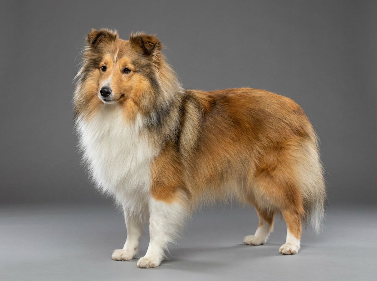
Corneal dystrophy is not among the five conditions documented for the Shetland Sheepdog, whose recorded eye disease is Collie Eye Anomaly. It is nonetheless a finding vets encounter across many breeds, and the association with thyroid disease gives it particular relevance here, since hypothyroidism is documented in this breed. Most Sheltie owners will never see it; those who do are usually looking at a benign finding with a useful diagnostic implication.
There is no documented corneal dystrophy predisposition in the Shetland Sheepdog. The genuine breed link runs through metabolism rather than through the eye itself. Hypothyroidism is documented here, and low thyroid hormone slows the clearance of circulating fats, which can then deposit in corneal tissue. That gives a Sheltie a plausible route to a corneal lipid deposit that is entirely secondary and entirely treatable at its source. The breed's own inherited eye condition, Collie Eye Anomaly, affects the vascular layer beneath the retina at the back of the eye and is unrelated to corneal clarity.
Environment contributes mainly through injury. A herding breed working through hedgerows and stubble collects corneal scratches, and a healed ulcer sometimes leaves mineral or scar deposit behind that looks like dystrophy but is not. Diet plays a modest role: a high-fat diet or frequent fatty treats can raise circulating lipids, which is why vets usually ask for a fasted blood sample when investigating a deposit. Chronic irritation from wind, dust or an inturning eyelash can also drive corneal changes over time.
Seek same-day care for any cloudy eye that is also painful: squinting, held shut, weeping heavily, red, or being pawed at. Pain plus a blue or diffuse haze across the whole cornea can mean glaucoma or inflammation inside the eye, both of which cost sight quickly. Book a routine appointment for a painless greyish or crystalline patch in both eyes, and expect the conversation to include thyroid and lipid bloodwork rather than eye drops. Ask for referral to a veterinary ophthalmologist if the deposit is large, central, changing, or if there is any question about how much vision is affected.
See all Shetland Sheepdog health problems, which breeds are prone to corneal dystrophy, or the full Shetland Sheepdog breed guide for temperament, exercise needs and ownership costs.
Bloodwork results come back within days. Where a thyroid cause is found and treated, changes in the corneal deposit are assessed over six to twelve months rather than weeks. Hereditary dystrophy with no underlying cause is monitored annually and often does not change for years.
A stable deposit, a dog with functional vision, and any underlying metabolic problem identified and treated. In many cases the eye finding never causes a single practical difficulty, and its whole value turns out to have been as a signpost to something else.
The single most useful question about a cloudy eye is where the cloudiness sits. These are the layers, from front to back, and what each typically means.
Lipid deposited in the cornea often reflects what is circulating in the blood. When a vet finds a corneal lipid deposit, particularly in an adult dog, a reasonable next step is to look for a reason why fats are elevated. Hypothyroidism is the classic candidate, because low thyroid hormone reduces the rate at which the body clears circulating lipids, and elevated blood fats can then deposit in tissue including the cornea.
This matters more in a Shetland Sheepdog than in most breeds, because hypothyroidism is one of the five conditions documented on the breed's health profile. A corneal deposit in a Sheltie is therefore a legitimate prompt to check thyroid function rather than an eccentric addition to the bill. The same bloodwork looks at other causes of raised lipids, and a fasted sample is usually requested because a recent fatty meal can raise the values on its own.
The practical payoff is worth stating plainly. The corneal deposit itself frequently needs no treatment. What the investigation may uncover is a treatable systemic condition that was otherwise going to be found later, once the dog had gained weight under a coat that hides it, become lethargic, and developed the recurring skin and coat problems that low thyroid function produces.
One more distinction. A hereditary corneal dystrophy, meaning deposit with no underlying metabolic cause, occurs in a number of breeds and needs no treatment at all beyond periodic monitoring. Distinguishing hereditary dystrophy from secondary lipid deposition is exactly what the bloodwork is for, and it is a good reason to accept the test rather than declining it as unnecessary.

Fionn was seven when his owner Catriona noticed what she described as glitter in both his eyes, a small crystalline oval in roughly the same position on each. He was not squinting and showed no sign of discomfort, so she mentioned it at his annual booster rather than booking specially. The vet identified corneal lipid deposits and recommended a fasted blood panel including thyroid function. Fionn's thyroid was underactive. In hindsight Catriona could list the signs she had attributed to age: he had gained two pounds she could not see under his coat, had been slower on walks all winter, and had had two ear infections that year. He was treated for the thyroid condition. The corneal deposits stopped enlarging and his energy came back over about three months.
Key takeaway: A painless eye finding can be the most visible sign of a systemic problem. Take the blood test that gets offered with it.
No. A cataract sits in the lens, deep inside the eye behind the pupil. Corneal dystrophy sits in the transparent dome at the very front. From a distance they can look similar, but they are different structures with entirely different implications.
Usually very little. Most corneal deposits are small and peripheral enough that the dog sees around them. A large central deposit can scatter light and reduce clarity, which is one reason for periodic ophthalmic reassessment rather than ignoring it.
Because corneal lipid deposits often reflect elevated blood fats, and hypothyroidism is a common cause of that. Since hypothyroidism is documented in the Shetland Sheepdog, the test is a reasonable and potentially valuable step.
There is no drop that dissolves an existing deposit. If an underlying metabolic cause is found and treated, some deposits reduce over months. Otherwise the approach is monitoring rather than intervention.
Photograph both eyes in the same light every few months from the same distance. Comparing images is far more reliable than comparing impressions, and it gives your vet something objective to look at.
When it comes with pain. Squinting, holding the eye shut, heavy tearing, redness, pawing at the face or shying from light means same-day veterinary care regardless of what the cloudiness looks like.
A greyish, silvery or crystalline patch within the clear front surface of the eye, usually in both eyes and usually painless.
Because raised blood fats deposit in the cornea, and hypothyroidism is a documented condition in this breed that raises them.
No, but a cloudy eye with pain always is. Squinting, tearing or rubbing means a same-day appointment whatever the appearance.
Photograph both eyes in the same light every few months from the same distance and compare the images rather than your memory.
The deposit itself usually needs none. Thyroid bloodwork and an ophthalmology consultation are modest one-off costs relative to the breed's monthly budget.
A one-page prep sheet for this condition: the signs, the questions to ask, what to get priced. Unlocks here.
You get: Vet visit prep sheet
No spam. One-click unsubscribe. See our privacy policy.
See a sample page before you decide.
Early access list
The free prep sheet covers the same-day warning signs, a symptom log and the questions to ask. We are considering a fuller kit that adds a cost worksheet for the usual tests and treatments (US price ranges with their sources) and a plain-English guide to ways of covering the bill. It is not built yet; this list tells us whether it is worth building.
One email, no spam, unsubscribe anytime. Information only, not veterinary advice.