Health
What is the difference between sclerosis and a cataract?
Sclerosis is a benign bluish haze of ageing that barely affects sight. A cataract is a white opacity in the lens that blocks light and does not reverse.

Quick answer
What is the difference between sclerosis and a cataract?
Sclerosis is a benign bluish haze of ageing that barely affects sight. A cataract is a white opacity in the lens that blocks light and does not reverse.
Most owners who notice cloudiness in an older dog's eyes are looking at something completely harmless. Telling the harmless version from the sight-threatening one is the whole job of this page.
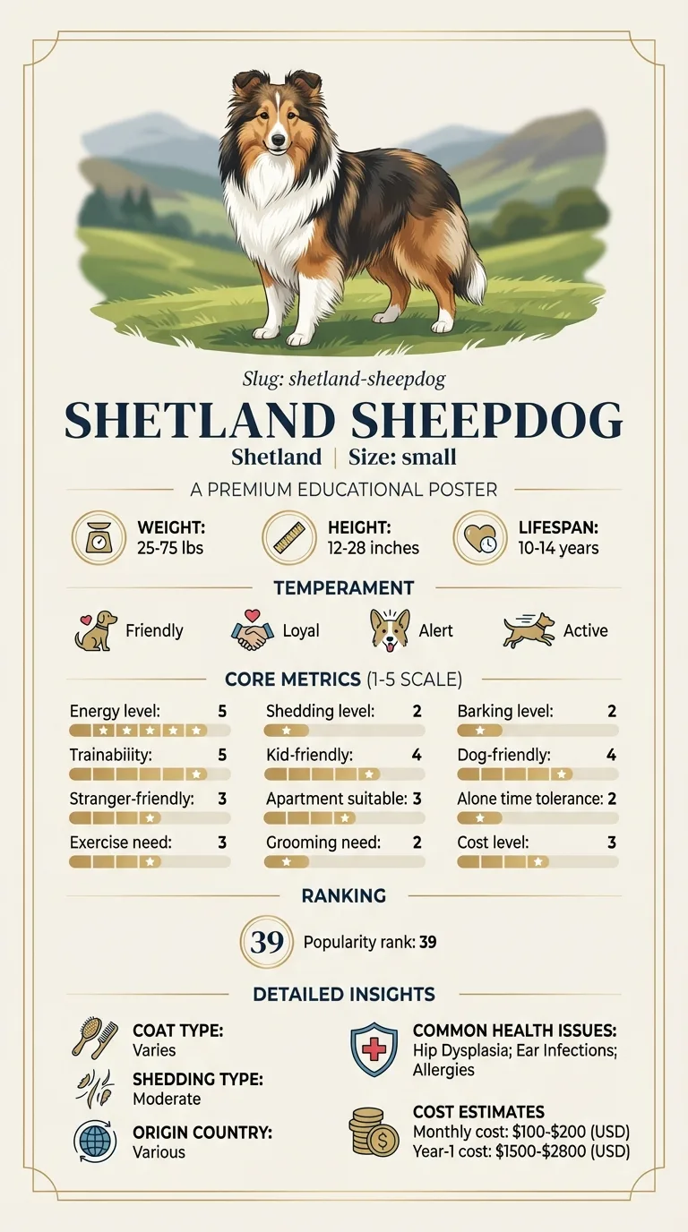
Owners of an ageing Sheltie who have noticed a bluish haze in both eyes. People whose young dog has developed a white patch in one eye. Anyone weighing whether cataract surgery makes sense for a dog who is otherwise coping.
This guide is not medical advice. If your dog shows pain, sudden behavior change, or worsening symptoms, consult a licensed veterinarian.
Some links may be affiliate links. We may earn a commission at no extra cost to you. We only suggest products we believe are helpful for dog owners.
We research and check what we publish, but we do not warrant that any figure, date, price, or statement is accurate, complete, or current, and we accept no liability for any loss, cost, injury, or other harm arising from the use of this site. Use it as one reference among several. You remain responsible for your own decisions and for your dog's care - confirm anything that matters with a licensed veterinarian or another qualified professional before you act on it. Full disclaimer.
A cataract is any opacity within the lens, ranging from a pinpoint speck that never affects vision to a complete white blockage that leaves the dog functionally blind in that eye. The change is structural and irreversible without surgery. What matters clinically is how much of the lens is involved, how fast it developed, whether the retina behind it is healthy, and whether the lens is provoking inflammation inside the eye.
Cataracts are not on the Shetland Sheepdog's documented health list, which records Collie Eye Anomaly as the breed's inherited eye condition. Cataracts do occur in individual Shelties, as they do in all breeds, and they become more likely in the later part of a 12 to 14 year lifespan. The far more common finding in an older Sheltie is benign nuclear sclerosis, which is frequently mistaken for a cataract by owners and occasionally by inexperienced observers.
The Shetland Sheepdog does not carry a documented cataract predisposition. Its inherited eye disease is Collie Eye Anomaly, which involves the vascular layer beneath the retina and is present from birth rather than developing later. That matters here in a specific way: a Sheltie with both conditions may have a lens problem in front of a retina that was never fully normal, and only a specialist examination separates how much of any vision loss belongs to which. Longevity of 12 to 14 years also gives ordinary age-related lens change plenty of time to appear.
Environment plays a limited role. Blunt trauma to the eye, whether from a branch, a thorn or a scuffle with another dog, can produce a cataract in the affected eye. Long-term inflammation inside the eye following an untreated injury does the same. Metabolic factors are more important than physical ones: a dog who develops diabetes may cloud over within weeks regardless of environment, and undiagnosed diabetes is often first suspected when an owner notices the eyes rather than the drinking.
Seek same-day help for a cloudy eye that is also red, painful, weeping, or being held shut, and for any sudden apparent loss of vision. Pain plus cloudiness can mean glaucoma or inflammation inside the eye, both of which damage sight quickly. Book promptly for eyes that have clouded noticeably over weeks rather than months, especially alongside increased thirst, more frequent urination or weight loss, since that combination suggests diabetes. Book a routine appointment for a gradual bluish haze in both eyes of an older dog, which is usually benign but is worth confirming with a dilated examination rather than assuming.
See all Shetland Sheepdog health problems, which breeds are prone to cataracts, or the full Shetland Sheepdog breed guide for temperament, exercise needs and ownership costs.
Diabetic cataracts can develop within weeks; inherited and age-related ones over months to years. Surgical assessment including retinal testing typically takes a few weeks to arrange. After surgery, intensive eye medication runs for several weeks and vision is usually functional within days.
For most Sheltie owners, success is a diagnosis of benign sclerosis and a dog who needs nothing at all. Where a true cataract exists, success is either restored functional vision after surgery or a comfortable, confident blind dog navigating a house that has been arranged around her.
Nuclear sclerosis is the normal age-related hardening of the lens centre. It is not a disease and barely affects vision. Distinguishing it from a cataract at home is imperfect, but these features help.
Cataracts arise by several routes. Inherited cataracts appear in specific breed lines, sometimes in dogs only a year or two old, and are usually bilateral. Diabetic cataracts are the most dramatic: high blood glucose draws water into the lens and can turn a clear eye opaque in a matter of weeks, sometimes in both eyes almost simultaneously. Trauma to the eye can produce a cataract in the injured eye alone, and long-standing inflammation inside the eye does the same. Age contributes too, though far less often than owners assume.
That causal split matters because a sudden cataract in an adult dog is a reason to test for diabetes rather than to accept as ageing. A dog who is drinking more, urinating more and losing weight while eating well, with rapidly clouding eyes, should be seen quickly.
Surgery genuinely works. A specialist removes the diseased lens and usually implants an artificial one, and functional vision is restored in a large majority of appropriately selected cases. Selection is the operative word. Candidates need a healthy retina, confirmed by electrical testing, since restoring the lens achieves nothing if the retina behind it has already degenerated. They need stable general health, controlled diabetes if relevant, and an owner able to manage an intensive eye medication schedule for weeks afterwards.
For a Shetland Sheepdog there is a specific reason to check the retina carefully. Collie Eye Anomaly is documented in this breed and affects the tissue layer underneath the retina. A dog with both a cataract and significant underlying eye disease may not be a surgical candidate, and finding that out before surgery rather than after is the point of the workup. The procedure itself requires general anesthesia, which makes the breed's documented MDR1 drug sensitivity relevant to the planning.

Nairn was ten and his owner Rosalind had noticed his eyes looking slightly milky over the summer. She assumed it was the same ageing haze her previous dog had developed. What made her book an appointment was unrelated: he had started emptying the water bowl twice a day and asking to go out in the night. Bloodwork showed diabetes. The cataracts had formed over roughly six weeks and were already dense. Nairn was stabilised on treatment, and once his glucose was controlled he was assessed by an ophthalmologist, whose retinal testing showed a healthy retina. He had surgery on both eyes four months later and regained functional vision. Rosalind's reflection was that the eyes had been the first sign and she had read them as the least alarming one.
Key takeaway: Eyes that cloud in weeks rather than years are a metabolic signal. Look at the water bowl before you blame the calendar.
Usually not. Nuclear sclerosis, the benign hardening of the lens with age, is far more common and affects vision very little. It looks bluish and translucent rather than white and opaque. A dilated examination settles it.
No. Various products are marketed for this and none reliably reverses lens opacity. Surgical removal is the only established treatment. Drops your vet prescribes are usually managing inflammation rather than the cataract itself.
Rapid bilateral clouding in an adult dog is strongly suggestive of diabetes and needs a prompt appointment with bloodwork. Look for increased thirst, more frequent urination and weight loss despite a good appetite alongside it.
Remarkably well, particularly when it develops slowly in familiar surroundings. Dogs navigate largely by scent and memory. The main adjustments are keeping furniture in fixed positions, using verbal cues and blocking stairs or water hazards.
Not usually in itself. It becomes painful if the lens material leaks and provokes inflammation inside the eye, or if it causes glaucoma. A red, squinting, tearing eye alongside a cataract is a same-day veterinary problem.
Where an inherited cataract is suspected, no. Eye certification by a specialist ophthalmologist is part of responsible breeding in this breed already, given that Collie Eye Anomaly is documented here.
Sclerosis is a benign bluish haze of ageing that barely affects sight. A cataract is a white opacity in the lens that blocks light and does not reverse.
Diabetes is the classic cause of rapid bilateral clouding. It warrants prompt bloodwork rather than waiting for the next check-up.
Yes, in well-selected cases, provided the retina behind the lens is healthy. Retinal testing is part of deciding whether surgery is worthwhile.
Keep furniture where it is, block stairs and ponds, and start using consistent verbal cues before sight is fully gone.
Specialist surgery with pre-operative testing runs to thousands, far above the breed's usual eighty to a hundred and fifty dollar monthly cost.
A one-page prep sheet for this condition: the signs, the questions to ask, what to get priced. Unlocks here.
You get: Vet visit prep sheet
No spam. One-click unsubscribe. See our privacy policy.
Preview page 1 before you decide.