Health
Is cherry eye common in Shelties?
No. It affects breeds with loose orbital tissue such as Bulldogs and Beagles and is not a recognised Shetland Sheepdog problem.

Quick answer
Is cherry eye common in Shelties?
No. It affects breeds with loose orbital tissue such as Bulldogs and Beagles and is not a recognised Shetland Sheepdog problem.
There is one rule about cherry eye that matters more than everything else written about it: the gland must be repositioned, never removed. Owners who understand that ask better questions than owners who do not.
Owners who have spotted something red at the inner corner of a Sheltie's eye and searched the most alarming term available. People whose dog has been offered gland removal and want to know whether that is acceptable. Anyone screening breeds for eye problems before buying.
This guide is not medical advice. If your dog shows pain, sudden behavior change, or worsening symptoms, consult a licensed veterinarian.
Some links may be affiliate links. We may earn a commission at no extra cost to you. We only suggest products we believe are helpful for dog owners.
We research and check what we publish, but we do not warrant that any figure, date, price, or statement is accurate, complete, or current, and we accept no liability for any loss, cost, injury, or other harm arising from the use of this site. Use it as one reference among several. You remain responsible for your own decisions and for your dog's care - confirm anything that matters with a licensed veterinarian or another qualified professional before you act on it. Full disclaimer.
Cherry eye is the forward displacement of the tear gland attached to the third eyelid, producing a smooth red or pink mass in the inner corner of the eye. It is usually painless at first but becomes irritated and inflamed if left, and the gland cannot function properly out of position. Its breed distribution is tightly linked to loose orbital and facial tissue, which places the Shetland Sheepdog well outside the affected group.
Rare to the point of being unexpected in this breed. Cherry eye does not appear on the Shetland Sheepdog's documented health list, which records Collie Eye Anomaly as its inherited eye condition. When a Sheltie owner sees something abnormal at the inner corner of the eye, the realistic explanations are conjunctivitis, an elevated third eyelid signalling pain or illness, or a foreign body lodged behind the membrane.
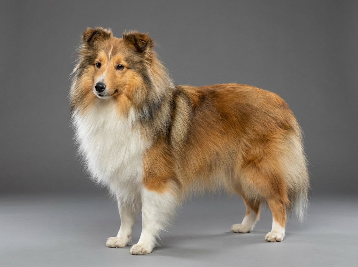
The relevant anatomy is the strength of the tissue anchoring the gland and the tightness of the orbit around it. The Shetland Sheepdog has a long, refined wedge-shaped head with an almond eye set obliquely and well supported, which is close to the opposite of the loose, shallow orbit that predisposes a Bulldog. Nothing in the breed's documented conditions relates to the third eyelid. What the breed does carry is Collie Eye Anomaly, a developmental abnormality of the tissue beneath the retina, which is a wholly separate structure at the back of the eye.
For a Sheltie, the environment supplies almost all the third eyelid problems that do occur. A dog working nose-first through hedgerows, stubble and long grass picks up grass seeds that lodge behind the membrane and cause sudden, intense one-sided irritation. Dust, wind and cleaning aerosols provoke conjunctival inflammation. Rubbing an already irritated eye against carpet or a paw makes any of it worse quickly, which is why an eye that is being pawed at needs attention the same day rather than the same week.
Seek same-day care for any eye your dog is holding shut, pawing at, or that is producing a sudden flood of tears, and for an eye that is red and painful or looks cloudy. Grass seeds and corneal scratches worsen by the hour, and pain in an eye is never something to observe overnight. Book promptly for a persistently visible third eyelid in an awake, alert dog, for a lump on the third eyelid in an older dog, or for repeated bouts of conjunctivitis, which often means an underlying allergy or an eyelid abnormality. If surgery on the third eyelid is ever proposed, ask explicitly whether the gland will be repositioned rather than removed.
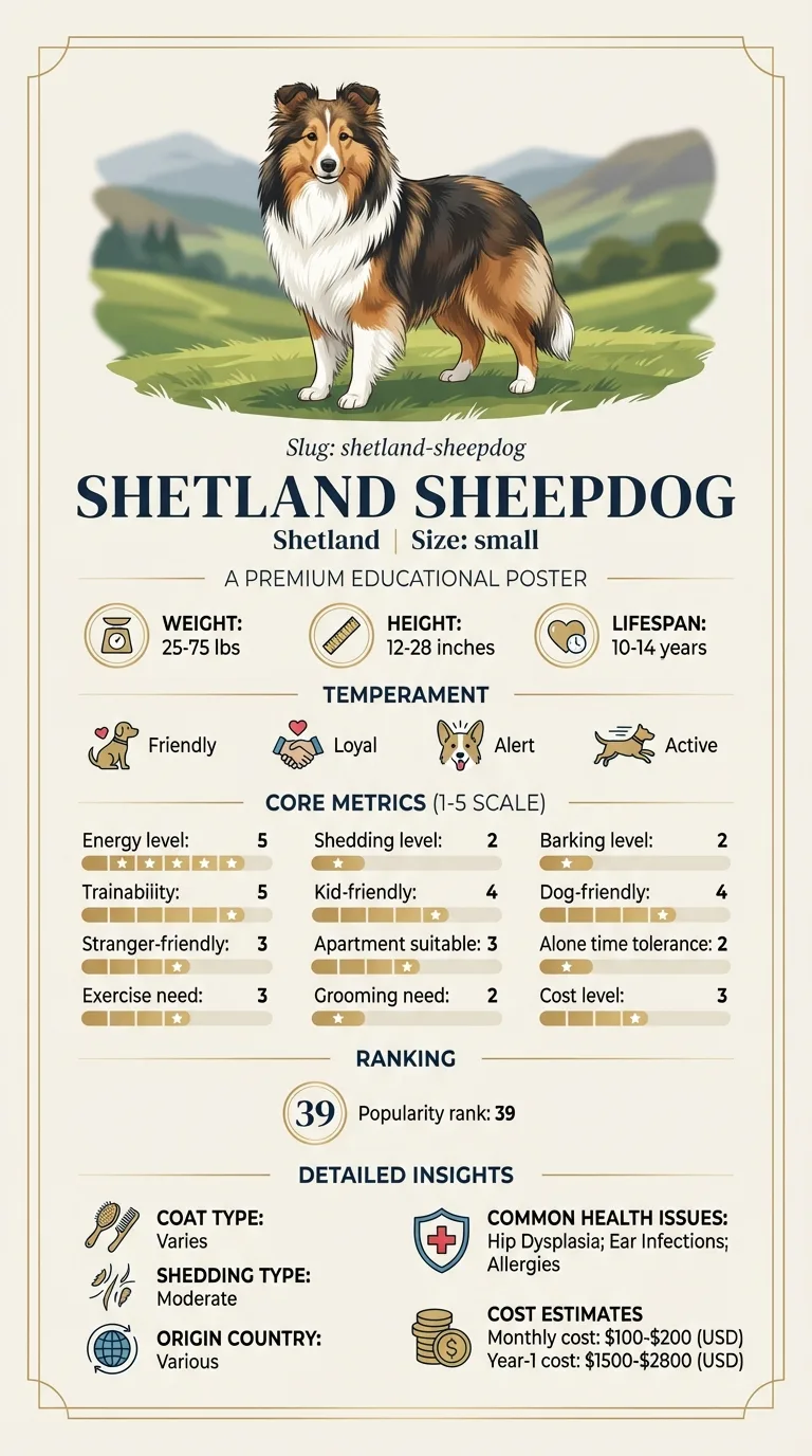
See all Shetland Sheepdog health problems, which breeds are prone to cherry eye, or the full Shetland Sheepdog breed guide for temperament, exercise needs and ownership costs.
Conjunctivitis usually settles in five to ten days with appropriate treatment. A grass seed is removed in one visit and the eye is comfortable within a day or two. Surgical gland repositioning, in the rare case it is needed, heals over two to three weeks with a recurrence rate that occasionally requires a second attempt.
A comfortable, clear, open eye with a third eyelid you rarely see. In this breed the realistic outcome for almost every red inner corner is a correctly identified irritation, treated quickly, with no long-term consequence at all.
Something visible at the inner corner of the eye is worth examining, but in this breed the differential list rarely starts with a prolapsed gland. These are the more likely explanations.
The gland of the third eyelid produces a large proportion of a dog's aqueous tears. For years the standard surgical approach to a prolapsed gland was to remove it, on the reasonable-looking grounds that a lump that will not stay put can simply be taken away. That practice has been abandoned by ophthalmologists, because the consequence turned up years later.
Dogs who lose that gland are substantially more likely to develop dry eye, a chronic condition where insufficient tear production leaves the cornea inflamed, thickened, pigmented and painful. It requires lifelong medication applied one to three times daily and, if poorly controlled, it damages vision. Trading a visible lump for a lifetime of drops and a deteriorating cornea is a poor bargain.
The accepted approach is replacement, most commonly by creating a pocket of conjunctival tissue that the gland is tucked into and secured within, so it returns to its correct position and keeps working. Recurrence is possible and a second procedure is sometimes needed, which is a considerably better problem than dry eye.
If you are ever offered gland removal for any dog, ask directly whether repositioning has been considered and whether referral to an ophthalmologist is appropriate. For a Shetland Sheepdog this conversation is unlikely to arise, but the surrounding principle applies broadly: eye surgery under general anesthesia in this breed should be planned with the documented MDR1 drug sensitivity known rather than assumed.

Whin came back from a walk through a barley stubble field with her left eye streaming and the inner corner looking red and swollen. Her owner Fergus searched online, found photographs of cherry eye and spent an evening reading about a condition his dog turned out not to have. At the appointment the following morning the vet applied a local anesthetic drop, lifted the third eyelid and removed a barley awn that had worked its way behind it. The redness was inflammation, not a prolapsed gland. Whin was comfortable within two days. Fergus now checks both eyes after every walk between June and September, which takes about fifteen seconds and has already caught a second seed.
Key takeaway: In a working breed, a red eye in late summer is a foreign body until proven otherwise. Check the eyes after the field, not after the streaming starts.
It is not a recognised problem in the breed. Cherry eye is associated with loose orbital and facial tissue, and the Sheltie has a tight, refined head with a well-anchored almond eye. The breed's documented eye condition is Collie Eye Anomaly instead.
A pale sliver at the inner corner in a relaxed or sleepy dog is normal anatomy. What is not normal is the membrane covering a significant part of the eye while your dog is awake and alert, which points to pain or illness.
Yes. Bilateral elevation often reflects a general problem such as dehydration, systemic illness or weight loss rather than an eye disease. It deserves a full examination rather than eye drops.
Do not attempt it. The tissue is delicate and easily damaged, and a gland pushed back without securing it simply prolapses again. A vet may reduce it temporarily to relieve irritation, but the definitive answer is surgical repositioning.
Pain is the divider. A dog holding an eye shut, tearing heavily, pawing at her face or shying from light needs same-day veterinary care. A calm dog with a small amount of visible pink tissue and no discomfort can wait for a routine appointment.
One-sided signs point to a local cause: a scratch on the cornea, a grass seed behind the third eyelid, or an inturning eyelash. All are treatable, and all get worse if the dog is left to rub at the eye for days.
No. It affects breeds with loose orbital tissue such as Bulldogs and Beagles and is not a recognised Shetland Sheepdog problem.
No. Removal greatly increases the risk of lifelong dry eye. The correct surgery repositions the gland so it keeps producing tears.
Usually pain, dehydration or general illness rather than a problem with the eyelid itself. It warrants a full examination.
Do not use leftover drops or human products. Rinsing with sterile saline is the limit, and a painful eye needs a vet the same day.
Repositioning surgery with anesthesia runs into several hundred dollars, above the breed's ordinary eighty to a hundred and fifty dollar monthly cost.
A one-page prep sheet for this condition: the signs, the questions to ask, what to get priced. Unlocks here.
You get: Vet visit prep sheet
No spam. One-click unsubscribe. See our privacy policy.
See a sample page before you decide.
Early access list
The free prep sheet covers the same-day warning signs, a symptom log and the questions to ask. We are considering a fuller kit that adds a cost worksheet for the usual tests and treatments (US price ranges with their sources) and a plain-English guide to ways of covering the bill. It is not built yet; this list tells us whether it is worth building.
One email, no spam, unsubscribe anytime. Information only, not veterinary advice.