Health
What does cherry eye look like?
A smooth, round, reddish lump suddenly appearing in the inner corner of the eye.

Quick answer
What does cherry eye look like?
A smooth, round, reddish lump suddenly appearing in the inner corner of the eye.
One day your Old English Sheepdog's eye looks completely normal, and the next there's an alarming red lump sitting in the corner. It looks far worse than it typically is.
This most often appears in younger dogs, and it catches owners off guard specifically because of how sudden and visually dramatic the change is, even though the underlying issue is a fairly well-understood, treatable condition.
This guide is not medical advice. If your dog shows pain, sudden behavior change, or worsening symptoms, consult a licensed veterinarian.
Some links may be affiliate links. We may earn a commission at no extra cost to you. We only suggest products we believe are helpful for dog owners.
We research and check what we publish, but we do not warrant that any figure, date, price, or statement is accurate, complete, or current, and we accept no liability for any loss, cost, injury, or other harm arising from the use of this site. Use it as one reference among several. You remain responsible for your own decisions and for your dog's care - confirm anything that matters with a licensed veterinarian or another qualified professional before you act on it. Full disclaimer.
Check this first
Cherry eye itself is uncomfortable but rarely an emergency in the truest sense, and many cases can be scheduled for surgery within a reasonable window rather than that same day. However, if the exposed gland becomes significantly swollen, discolored, or the eye shows signs of pain, heavy discharge, or the cornea looks affected, prompt veterinary attention is warranted rather than waiting for a routine appointment slot.
A good rule of thumb: if the lump appeared today and your dog is otherwise comfortable, a call to your vet within a day or two to schedule an exam is reasonable. If the eye looks increasingly irritated, painful, or your dog is pawing at it persistently, ask for a sooner appointment.
If any of these apply, call your vet or an emergency clinic now. The rest of this page is for the cases that are not an emergency.
Cherry eye is the prolapse of the third eyelid's tear-producing gland, appearing suddenly as a reddish lump in the inner corner of the eye. It's a structural, connective-tissue issue rather than an infection or injury, and it's generally treated surgically with a technique that preserves the gland rather than removing it.
Cherry eye occurs across many breeds and isn't unique to Old English Sheepdogs, but it's a recognizable, well-understood condition that owners of any breed with third eyelid anatomy, including this one, may encounter, most often in younger dogs.
Cherry eye results from weakness in the connective tissue anchoring the third eyelid's gland in place, allowing it to prolapse forward. It isn't specifically tied to this breed's documented health issues, but the underlying anatomy involved is present in all dogs, and any individual dog's connective tissue can be more or less prone to it.
Physical irritation, such as rubbing or pawing at the eye, can sometimes trigger or worsen a prolapse once the underlying tissue weakness is present, though many cases appear without any clear external trigger.
Schedule a vet visit within a day or two of noticing the lump. Seek prompt attention sooner if the eye appears increasingly swollen, painful, or your dog is persistently pawing at it.
Cherry eye responds much better to prompt, correct treatment than to improvised home approaches.
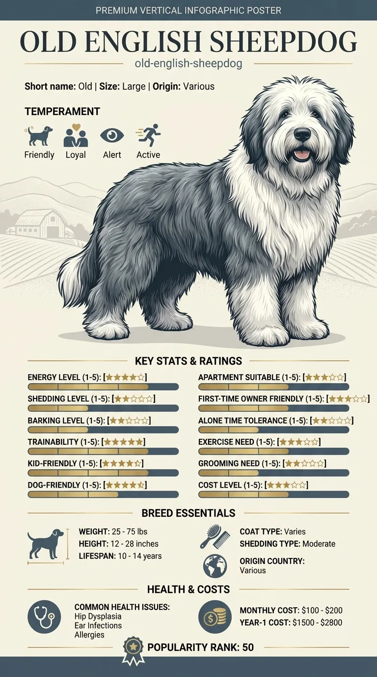
See all OLD English Sheepdog health problems, which breeds are prone to cherry eye, or the full OLD English Sheepdog breed guide for temperament, exercise needs and ownership costs.
Once diagnosed, surgery is often scheduled within a week or two, and recovery typically takes about one to two weeks with an e-collar to prevent rubbing at the healing area.
Success looks like the gland staying in position after surgical correction, with normal tear production preserved and no recurrence, though watching the other eye for a future prolapse remains worthwhile.
If you think your Old has cherry eye, the plan is three steps: write down what you have seen and when it started, book a veterinary appointment rather than waiting for the next flare, and take video of the behaviour or symptom before you go — the thing you are worried about rarely happens in the consulting room. Screening in the parents covers hip dysplasia, cataracts, hypothyroidism.
What the vet visit should produce is a diagnosis and a written plan, not just reassurance: what is being ruled out, what the monitoring interval is, and which signs mean you come back sooner. Ask what the treatment costs across a year rather than per visit, because that is the number that decides whether you are managing this condition or reacting to it.
Between appointments, keep a short log — dates, what you saw, what changed. It is the single most useful thing an owner brings to a follow-up, and for cherry eye it is often what separates a clear pattern from a guess.
Pippin, a two-year-old Old English Sheepdog, seemed completely normal at bedtime. The next morning, his owner noticed a smooth red lump in the corner of one eye and panicked, assuming it was a tumor or injury. A same-week vet visit confirmed cherry eye, and the vet recommended the gland-tacking surgical technique rather than removal. Pippin recovered fully within about two weeks, wearing a cone to prevent rubbing at the site. Eight months later, the second eye prolapsed as well, and the same procedure resolved it just as effectively.
Key takeaway: Cherry eye looks alarming, but it's a well-understood, treatable structural issue. Acting promptly, and asking about gland-preserving surgery specifically, gives the best long-term outcome.
It's the prolapse of a tear-producing gland normally hidden behind the third eyelid, in the inner corner of the eye. When the connective tissue holding it in place weakens, the gland pops forward and becomes visible as a reddish lump.
It's more often described as irritating than acutely painful, though the exposed gland can become swollen or irritated over time, especially if a dog rubs at it or if treatment is delayed.
It's uncommon for it to resolve without treatment. Most cases require a vet visit and typically a surgical procedure to correct it properly.
The current standard approach is a surgical technique that tacks the gland back into its normal position, preserving its function, rather than removing it outright. Removal is generally avoided when possible since the gland contributes meaningfully to tear production.
Removing the gland can increase the risk of dry eye later in life, since it plays a role in tear production. This is why the tacking approach is generally preferred when it's a viable option.
Typically a few hundred to around a thousand dollars per eye, depending on the practice, region, and whether both eyes are affected.
Yes, it can occur in one eye and later in the other, since the underlying weakness in connective tissue isn't necessarily limited to one side.
A smooth, round, reddish lump suddenly appearing in the inner corner of the eye.
Uncommonly; most cases need a vet visit and typically surgery to correct properly.
Tacking it back into position is generally preferred to preserve tear production and reduce future dry-eye risk.
Usually not urgent same-day unless the eye looks increasingly painful or irritated; a vet visit within a day or two is reasonable otherwise.
Often a few hundred to around a thousand dollars per eye for the surgical correction.
A one-page prep sheet for this condition: the signs, the questions to ask, what to get priced. Unlocks here.
You get: Vet visit prep sheet
No spam. One-click unsubscribe. See our privacy policy.
Preview page 1 before you decide.