Health
What part of the eye does corneal dystrophy affect?
The cornea, the clear front surface of the eye, not the lens inside it, which is what a cataract affects.

Quick answer
What part of the eye does corneal dystrophy affect?
The cornea, the clear front surface of the eye, not the lens inside it, which is what a cataract affects.
Two different conditions can make an eye look hazy, and telling them apart matters because they behave completely differently. Corneal dystrophy usually stays superficial, painless and stable; a cataract can progress toward significant vision loss. A vet exam, not an owner's naked eye, is what actually distinguishes them.
Owners who notice a cloudy patch on the surface of their dog's eye and assume it must be a cataract, missing that a different, often less concerning condition could be responsible.
This guide is not medical advice. If your dog shows pain, sudden behavior change, or worsening symptoms, consult a licensed veterinarian.
Some links may be affiliate links. We may earn a commission at no extra cost to you. We only suggest products we believe are helpful for dog owners.
Corneal dystrophy is an inherited condition causing lipid or mineral deposits to build up within the cornea, the eye's clear front surface, producing a cloudy, often ring-shaped appearance. It is distinct from cataracts, which affect the lens, and is typically painless and stable rather than progressive.
Corneal dystrophy is a recognized condition across several dog breeds and is not uncommon as an incidental finding during routine eye exams, though its overall frequency specifically in Saint Bernards has not been extensively documented.
Corneal dystrophy arises from inherited genetic factors affecting how lipid or mineral material is processed and deposited within corneal tissue. There is no strong documentation of this being uniquely elevated in Saint Bernards, though it remains a recognized possibility as it is across many breeds.
There is little established environmental influence on typical corneal dystrophy, since it is primarily a genetic condition. Age can play a role in how visible existing deposits become over time, even when the underlying process remains stable.
See a vet for any new corneal cloudiness to confirm the diagnosis and rule out other conditions. Treat as more urgent any redness, squinting, apparent pain, or a rapidly changing cloudy patch, since these suggest a different, potentially more serious corneal problem rather than typical stable dystrophy.
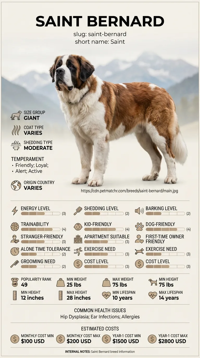
See all Saint Bernard health problems, which breeds are prone to corneal dystrophy, or the full Saint Bernard breed guide for temperament, exercise needs and ownership costs.
Diagnosis typically takes a single vet visit with a thorough eye exam. Most cases require only periodic monitoring rather than active treatment, and stability is generally assessed over months to years of rechecks.
Success is a confirmed, accurate diagnosis distinguishing this condition from more concerning eye problems, with periodic monitoring confirming the finding stays stable over time and does not meaningfully affect vision.
The cornea is the eye's outermost clear layer, and corneal dystrophy typically shows up as distinct, often symmetrical, grayish-white or silvery deposits within it, frequently in a ring or oval pattern. Because these deposits sit in the front layers of the eye, they are usually visible without any special equipment, but a vet exam with magnification and specific staining tests is what actually confirms the diagnosis and rules out other corneal conditions with a similar appearance.
Unlike a cataract, corneal dystrophy in its typical form does not usually progress to significant vision loss and is generally not painful. It can, in some cases, remain essentially stable for years once diagnosed, which is part of why an accurate diagnosis matters — mistaking it for a progressive, vision-threatening cataract can cause unnecessary worry, while mistaking a true cataract for this more benign condition risks missing a genuinely progressive problem.
Most corneal dystrophy cases stay quiet for years, but the exceptions above are exactly the signs that separate a stable, cosmetic finding from something that needs a closer look.

If you think your Saint has corneal dystrophy, the plan is three steps: write down what you have seen and when it started, book a veterinary appointment rather than waiting for the next flare, and take video of the behaviour or symptom before you go — the thing you are worried about rarely happens in the consulting room. Screening in the parents covers hip dysplasia, elbow dysplasia, dilated cardiomyopathy.
What the vet visit should produce is a diagnosis and a written plan, not just reassurance: what is being ruled out, what the monitoring interval is, and which signs mean you come back sooner. Ask what the treatment costs across a year rather than per visit, because that is the number that decides whether you are managing this condition or reacting to it.
Between appointments, keep a short log — dates, what you saw, what changed. It is the single most useful thing an owner brings to a follow-up, and for corneal dystrophy it is often what separates a clear pattern from a guess.
Nova, a five-year-old Saint Bernard, was found to have a faint, symmetrical grayish ring in both corneas during a routine wellness exam, something her owner had never noticed at home. Concerned it might be an early cataract, her owner asked for a referral to confirm. A veterinary ophthalmologist confirmed corneal dystrophy rather than a cataract, noting the deposits sat in the front corneal layers rather than the lens. Nova's vision was unaffected, and annual rechecks over the following three years showed no meaningful change in the pattern, confirming it as the stable, cosmetic finding it appeared to be from the start.
Key takeaway: The distinction between cornea and lens made all the difference to Nova's outlook — getting the right diagnosis, rather than assuming the worse-case explanation, spared her family years of unnecessary worry.
No. Corneal dystrophy affects the clear front surface of the eye, while a cataract clouds the lens deep inside it. They can look superficially similar but are different conditions with different outlooks.
Typically not. Most corneal dystrophy cases are painless and do not significantly affect vision, though any accompanying redness, squinting or apparent discomfort should be evaluated promptly.
Many cases remain fairly stable for years once diagnosed, though this varies by individual, which is why periodic rechecks are worthwhile to confirm stability.
Often no active treatment is needed beyond monitoring, since most cases do not progress to significant vision loss or discomfort. Your vet will advise based on your dog's specific presentation.
A vet exam with magnification, often alongside a fluorescein stain test to rule out other corneal conditions like ulcers, is generally used to confirm the diagnosis.
Typical corneal dystrophy usually does not progress to significant vision loss, unlike more severe or progressive corneal or lens conditions, though any change in vision should always be evaluated.
The cornea, the clear front surface of the eye, not the lens inside it, which is what a cataract affects.
Typically not — most cases are painless and do not significantly affect vision.
Many cases stay fairly stable for years, though periodic vet rechecks help confirm this for an individual dog.
You generally can't reliably by eye alone — a vet exam is needed to distinguish the two conditions accurately.
Often not — monitoring is common, since most cases remain stable without significant progression.
A one-page prep sheet for this condition: the signs, the questions to ask, what to get priced. Unlocks here.
You get: Vet visit prep sheet
No spam. One-click unsubscribe. See our privacy policy.
Preview page 1 before you decide.