Health
What does corneal dystrophy look like?
A faint silvery, crystalline, or hazy patch on the clear surface of the eye, often circular or oval in shape.

Quick answer
What does corneal dystrophy look like?
A faint silvery, crystalline, or hazy patch on the clear surface of the eye, often circular or oval in shape.
It often shows up as a faint, silvery or crystalline haze on the surface of the eye rather than deep inside it — subtle enough that many owners don't notice it until a vet points it out during a routine exam.
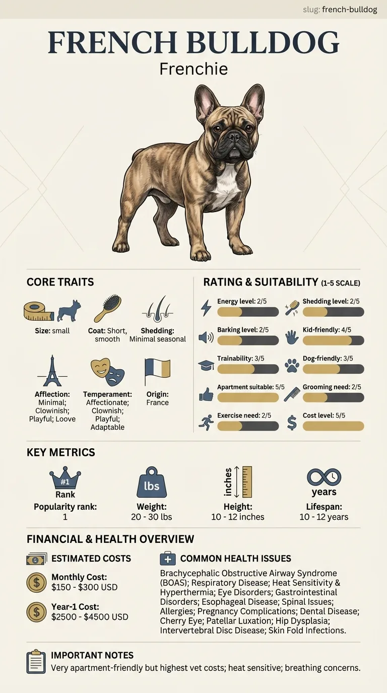
Owners who are meticulous about eye checkups are often the ones who catch this early, since in its mild form it produces no obvious symptoms at all. It becomes a bigger deal for owners of dogs where the deposits are dense enough to genuinely blur vision or cause the surface to break down.
This guide is not medical advice. If your dog shows pain, sudden behavior change, or worsening symptoms, consult a licensed veterinarian.
Some links may be affiliate links. We may earn a commission at no extra cost to you. We only suggest products we believe are helpful for dog owners.
We research and check what we publish, but we do not warrant that any figure, date, price, or statement is accurate, complete, or current, and we accept no liability for any loss, cost, injury, or other harm arising from the use of this site. Use it as one reference among several. You remain responsible for your own decisions and for your dog's care - confirm anything that matters with a licensed veterinarian or another qualified professional before you act on it. Full disclaimer.
Check this first
In a small number of dogs, the deposits weaken the surface layer of the cornea enough that it erodes, exposing a raw area underneath. That's when a previously painless condition turns painful fast — sudden squinting, redness, excess tearing, or pawing at the eye are signs the surface has broken down.
An eroded cornea is a same-day vet problem, not a wait-and-watch one, because an exposed cornea is vulnerable to infection and can scar in ways that permanently affect vision.
Outside of that erosion scenario, corneal dystrophy on its own rarely qualifies as an emergency. The distinction that matters is pain: a quiet, comfortable eye with visible haze is a monitoring situation; a red, squinting, watering eye is not.
If any of these apply, call your vet or an emergency clinic now. The rest of this page is for the cases that are not an emergency.
Corneal dystrophy refers to an inherited buildup of lipid or mineral material within the layers of the cornea — the clear, dome-shaped surface at the front of the eye. Unlike a cataract, it isn't inside the lens, and unlike an ulcer, it isn't caused by injury or infection. It typically appears as a stable or slowly changing hazy patch, most often noticed by a vet during a routine eye exam rather than by the owner at home.
Corneal dystrophy is diagnosed less frequently than conditions like cherry eye or cataracts in this breed, but it's recognized often enough in veterinary ophthalmology literature on brachycephalic breeds to be worth knowing about, particularly since it's usually silent until specifically looked for.
The tendency toward corneal deposits appears to run in certain lines, which is why it's classified as a breed-associated dystrophy rather than a random individual finding. It isn't caused by the flat face or breathing anatomy the way some other French Bulldog eye issues are — this one sits more purely in the genetics of the corneal tissue itself.
Environmental factors play a smaller role here than in many eye conditions. Chronic low-grade irritation from dust, dry air, or frequent eye rubbing could theoretically contribute to surface changes over time, but the core process is inherited rather than driven by the dog's surroundings.
Reach out promptly if a previously painless, stable eye suddenly becomes red, squinty, watery, or clearly bothers the dog — that combination suggests the corneal surface may have broken down and needs same-day evaluation rather than a routine appointment.
Because this condition is often silent, most mistakes come from either overreacting or under-monitoring.
See all French Bulldog health problems, which breeds are prone to corneal dystrophy, or the full French Bulldog breed guide for temperament, exercise needs and ownership costs.
Most corneal dystrophy is diagnosed incidentally and then followed with rechecks every six to twelve months indefinitely, since it's a lifelong, usually stable finding rather than something that resolves.
Success looks like a stable or very slowly changing deposit that never meaningfully affects vision or comfort — essentially, a finding that gets tracked but never becomes a crisis.
Piper, a four-year-old Frenchie, went in for a routine annual exam with no eye complaints at all. Her vet noted a faint, silvery, oval haze on the surface of her right eye during a slit-lamp check and diagnosed early corneal dystrophy. Piper's owner had genuinely never noticed anything unusual at home. With no pain or vision impact, the plan was simply to recheck in six months. Two years later, the deposit looks almost identical to that first exam. Piper's owner now asks specifically for a corneal check at every annual visit, since the finding was easy to miss and would have gone unnoticed without a targeted look.
Key takeaway: A silent condition still deserves a plan — regular monitoring turned an easy-to-miss diagnosis into a manageable, low-drama part of Piper's checkups.
No. A cataract clouds the lens deep inside the eye, while corneal dystrophy deposits build up in the clear window at the very front of the eye. They can look superficially similar to an owner but are entirely different structures and conditions.
Most cases stay mild and don't meaningfully affect vision. In a smaller number of dogs, dense deposits or resulting erosions can blur vision in the affected area of the eye, though total blindness from this condition alone is uncommon.
Typically not, as long as the corneal surface stays intact. It becomes painful only if the deposits cause the surface layer to break down, which shows up as sudden squinting, redness, and tearing.
There's no treatment that removes the deposits themselves in most cases. Management focuses on monitoring for progression and treating any resulting surface erosion promptly if it occurs.
Some forms are believed to have a genetic component, which is part of why it's tracked in breed health screening. If you're considering breeding a diagnosed dog, discuss the specific finding with your vet first.
A vet or veterinary ophthalmologist identifies it during a slit-lamp eye exam, often as an incidental finding during a routine checkup rather than something the owner brought the dog in for.
A general vet can usually identify and monitor it. A referral to a veterinary ophthalmologist is more useful if the deposits are dense, appear to be progressing, or the surface has started to erode.
A faint silvery, crystalline, or hazy patch on the clear surface of the eye, often circular or oval in shape.
Usually not, unless the deposits cause the corneal surface to break down, which then causes sudden pain and redness.
No cure removes the deposits; most cases are simply monitored over time for stability or progression.
It can, since some forms are believed to be hereditary — worth discussing with a vet before breeding an affected dog.
Roughly once or twice a year is typical for a stable finding, more often if deposits appear to be increasing.
A one-page prep sheet for this condition: the signs, the questions to ask, what to get priced. Unlocks here.
You get: Vet visit prep sheet
No spam. One-click unsubscribe. See our privacy policy.
Preview page 1 before you decide.