Health
What is the main eye disease in Shelties?
Collie Eye Anomaly, an inherited developmental defect of the tissue behind the retina. It is the breed's headline documented condition.

Quick answer
What is the main eye disease in Shelties?
Collie Eye Anomaly, an inherited developmental defect of the tissue behind the retina. It is the breed's headline documented condition.
Most affected Shelties see perfectly well their whole lives. A minority go blind, and the examination that separates them has to happen in a narrow window in early puppyhood.
Puppy buyers trying to interpret a breeder's eye certificate, and owners of adult Shelties wondering whether the annual eye examination they keep being offered is worth the money.
This guide is not medical advice. If your dog shows pain, sudden behavior change, or worsening symptoms, consult a licensed veterinarian.
Some links may be affiliate links. We may earn a commission at no extra cost to you. We only suggest products we believe are helpful for dog owners.
We research and check what we publish, but we do not warrant that any figure, date, price, or statement is accurate, complete, or current, and we accept no liability for any loss, cost, injury, or other harm arising from the use of this site. Use it as one reference among several. You remain responsible for your own decisions and for your dog's care - confirm anything that matters with a licensed veterinarian or another qualified professional before you act on it. Full disclaimer.
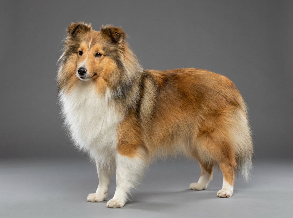
Eye disease in Shetland Sheepdogs is dominated by Collie Eye Anomaly, an inherited failure of the choroid to develop properly behind the retina. Most affected dogs have a mild form that never affects vision. A minority carry colobomas that can lead to retinal detachment and blindness, sometimes early in life. For owners the practical experience is paperwork and screening: a breeder's certificate at eight weeks, then annual examinations that mostly find nothing.
Collie Eye Anomaly is common in the rough-coated collie family and is documented for the Shetland Sheepdog. Mild findings on examination are far more frequent than colobomas, and vision-threatening disease affects a small proportion of affected dogs. Being affected and being at risk of blindness are quite different statements.
This is an inherited developmental condition specific to the collie group, and it is the first entry on the Shetland Sheepdog's documented health list alongside MDR1 drug sensitivity, dermatomyositis, hypothyroidism and patellar luxation. Because it is recessive and widespread in the breed's gene pool, it cannot simply be bred out in a generation, which is why examination and DNA testing exist. The breed's other health entries intersect here indirectly: an eye examination under sedation, or surgery on an eye, means MDR1 status has to be established beforehand.
Nothing an owner does causes Collie Eye Anomaly, and no diet, supplement or lighting condition influences it. What environment affects is the consequences and the detection. Head trauma can precipitate detachment in an eye with a coloboma, so a dog with that finding is worth protecting from high-impact activity. And a household that keeps furniture in fixed positions makes an enormous practical difference to a dog whose vision is failing, which is a genuine environmental intervention even though it is not a medical one.
Go immediately, treating it as an emergency: a sudden loss of vision, an eye that is red, cloudy, bulging or obviously painful, an eye the dog is holding shut or rubbing hard, or any injury to the eye. Glaucoma and retinal detachment are time-critical, and an acutely painful eye can be lost within a day. Book within days: bumping into things, new hesitancy in the dark, dilated pupils that do not respond to light, or a change in the appearance of either eye. Book annually: a screening examination with an ophthalmologist for any Shetland Sheepdog, and more often if a coloboma has been identified.
See all Shetland Sheepdog health problems, which breeds are prone to ocular disease, or the full Shetland Sheepdog breed guide for temperament, exercise needs and ownership costs.
Severity is fixed at birth and identified in one examination at six to eight weeks, so the diagnostic timeline is short. For dogs with mild findings, expect nothing to change for the whole of a twelve to fourteen year life. For dogs with colobomas, detachment risk is highest in the first couple of years but is never zero, which is why annual screening continues. Adaptation to vision loss, when it occurs, generally takes a few weeks in a familiar home.
Success for most Sheltie owners is a stack of annual reports that all say the same thing. For a dog with a coloboma it is reaching old age with the retina still attached. And for a dog that does lose vision, success is a dog that still runs the same route in the same garden with confidence, which is achievable far more often than owners expect from a breed this intelligent.
During development, the tissue layer that nourishes the retina, called the choroid, fails to form properly. The minimum finding is choroidal hypoplasia, a pale, thin patch visible to an ophthalmologist looking into the back of the eye. On its own it causes no vision problem at all, and the dog lives a completely normal life.
The problem is that the same developmental failure can go further. A coloboma is a pit or hole where the tissue at the optic disc did not close. Colobomas can lead to retinal detachment, and detached retinas mean blindness. Bleeding inside the eye is the third and least common consequence.
So the disease is best understood as a spectrum with a wide, harmless base and a narrow, serious tip. Roughly the great majority of affected dogs sit at the mild end. A minority have colobomas, and of those a smaller number lose vision, sometimes suddenly and sometimes in the first year or two of life.
The severity is determined at birth. It does not progress in the sense that a mild dog becomes a severe one; what can happen is that a dog with a coloboma suffers a detachment later. This is why a mild finding is genuinely reassuring and a coloboma genuinely is not.
Here is the fact that governs everything about screening in this breed. As a Sheltie puppy grows, pigment develops at the back of the eye and can mask a mild choroidal defect completely. A dog that was clearly abnormal at six weeks can examine as normal at six months.
Breeders call this a go normal, and it is not a recovery. The defect is still there and the dog will still pass the gene to its puppies. It has simply become invisible.
That is why litters are examined between about six and eight weeks, when the pigment has not yet developed and the finding is still visible. An examination done later can miss mild cases entirely, which is fine for predicting your own dog's vision and useless for breeding decisions.
The modern complement to this is a DNA test for the variant associated with the condition, which can be done at any age and does not care about pigment. It reports a dog as clear, carrier or affected. It tells you about inheritance rather than severity, so it does not replace the ophthalmologist's examination, which is what identifies whether a coloboma is present. The two answer different questions and a careful breeder uses both.
For a pet owner the practical takeaway is simple. Ask for the litter examination certificate with its date. If the date is after ten weeks, ask why.

If you think your Shetland has ocular disease, the plan is three steps: write down what you have seen and when it started, book a veterinary appointment rather than waiting for the next flare, and take video of the behaviour or symptom before you go — the thing you are worried about rarely happens in the consulting room. Screening in the parents covers collie eye anomaly, mdr1 drug sensitivity, dermatomyositis.
What the vet visit should produce is a diagnosis and a written plan, not just reassurance: what is being ruled out, what the monitoring interval is, and which signs mean you come back sooner. Ask what the treatment costs across a year rather than per visit, because that is the number that decides whether you are managing this condition or reacting to it.
Between appointments, keep a short log — dates, what you saw, what changed. It is the single most useful thing an owner brings to a follow-up, and for ocular disease it is often what separates a clear pattern from a guess.
Callie was looking at two Sheltie litters. Both breeders produced eye certificates. She almost took the first, until a Sheltie club member she had met at a show suggested she check the dates. The first litter had been examined at fourteen weeks. Every puppy was recorded as clear. The second had been examined at seven weeks, and two of the five puppies were recorded as affected with mild choroidal hypoplasia, one of which was the puppy Callie liked. On the face of it the first litter looked better. In fact the second breeder had done the examination properly, in the window where mild findings are still visible, and had reported the results honestly. The first breeder had, deliberately or not, screened at an age where mild cases go normal and disappear. Callie bought the affected puppy, whose ophthalmologist confirmed no coloboma and normal vision. That dog is now nine, sees perfectly, and has had an annual screening every year of her life.
Key takeaway: In this breed the date on the eye certificate matters as much as the result. A clear finding after ten weeks may simply mean the examination happened too late to see anything, and an honest affected result from a seven-week examination is often the better sign.
Probably not. The great majority of affected dogs have only mild choroidal hypoplasia and see normally for life. Vision loss occurs where colobomas lead to retinal detachment, which affects a small minority. What determines your dog's risk is what an ophthalmologist saw at the back of the eye, not simply whether it is affected.
A puppy with mild Collie Eye Anomaly whose developing pigment masks the finding, so a later examination appears clear. The dog is still affected genetically and still passes it on. This is the entire reason litters are examined at six to eight weeks rather than later.
For a pet dog, the breeder's eye examination usually tells you what you need. For anyone breeding, both are worth having: the DNA test establishes clear, carrier or affected status regardless of age, and the ophthalmologist's examination identifies whether the eye actually contains a coloboma.
They are worth more in this breed than in most. An annual screening examination catches retinal detachment risk, cataract, and progressive retinal atrophy at a stage where you can plan rather than react. In a dog with a known coloboma it is not optional.
Watch behaviour rather than eyes. Bumping into things in unfamiliar places, hesitancy on stairs, reluctance to go out at dusk, startling when approached, and a sudden loss of confidence in a previously bold dog are all more informative than looking at the eye itself.
Yes. Sudden vision loss, a painful eye, a cloudy or bulging eye, or an eye the dog is holding shut all need same-day veterinary attention. Retinal detachment, glaucoma and eye trauma are all time-sensitive, and delay in an acutely painful eye can cost the eye itself.
Answer their questions honestly. Be aware that a finding noted on a breeder's puppy examination predates any policy you buy and may be treated as pre-existing, which can exclude a large category of future claims. Obtain the certificate before you choose a policy rather than afterwards.
Collie Eye Anomaly, an inherited developmental defect of the tissue behind the retina. It is the breed's headline documented condition.
Between about six and eight weeks, before developing pigment can mask mild findings. Later examinations can miss affected dogs entirely.
Usually not at all. Choroidal hypoplasia on its own is a cosmetic finding on examination and most affected dogs see normally throughout life.
A screening examination by a veterinary ophthalmologist typically runs in the low hundreds, which is a modest annual addition for this breed.
Yes, remarkably so, provided furniture stays where it is. This breed's intelligence and trainability make verbal cue work very effective.
A one-page prep sheet for this condition: the signs, the questions to ask, what to get priced. Unlocks here.
You get: Vet visit prep sheet
No spam. One-click unsubscribe. See our privacy policy.
See a sample page before you decide.
Early access list
The free prep sheet covers the same-day warning signs, a symptom log and the questions to ask. We are considering a fuller kit that adds a cost worksheet for the usual tests and treatments (US price ranges with their sources) and a plain-English guide to ways of covering the bill. It is not built yet; this list tells us whether it is worth building.
One email, no spam, unsubscribe anytime. Information only, not veterinary advice.