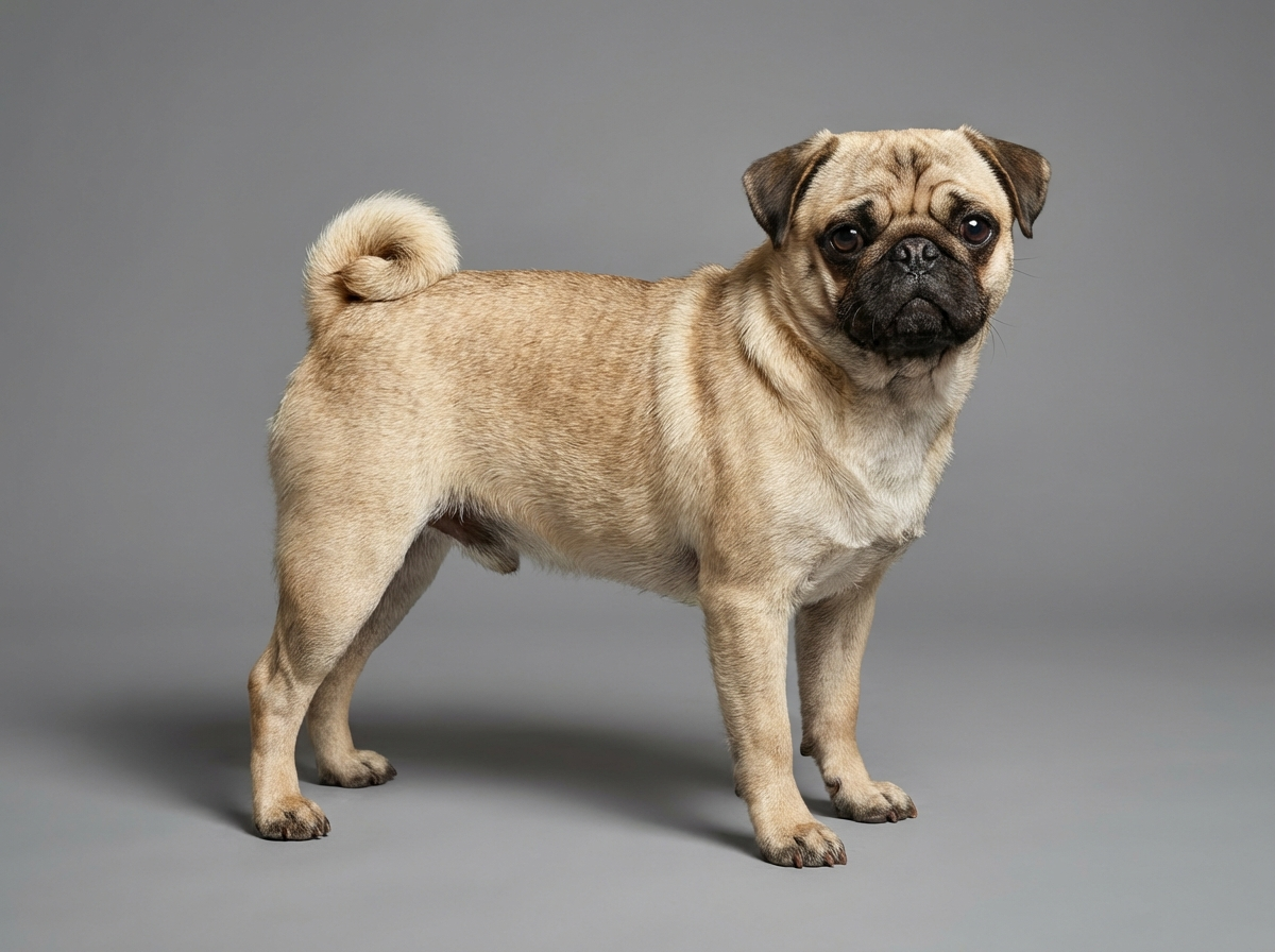
Health
What does cherry eye look like?
A smooth, reddish-pink bulge at the inner corner of the eye, often appearing suddenly.

Quick answer
What does cherry eye look like?
A smooth, reddish-pink bulge at the inner corner of the eye, often appearing suddenly.
The bulge looks dramatic, but cherry eye is more of an urgent nuisance than a crisis.
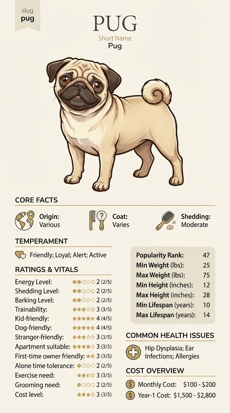
This most often affects young Pugs, typically under two years old, since weakened connective tissue in this gland tends to show up early in life rather than developing gradually with age the way many other eye conditions do.
This guide is not medical advice. If your dog shows pain, sudden behavior change, or worsening symptoms, consult a licensed veterinarian.
Some links may be affiliate links. We may earn a commission at no extra cost to you. We only suggest products we believe are helpful for dog owners.
Cherry eye occurs when weakened connective tissue allows the third eyelid's tear-producing gland to flip outward, appearing as a reddish bulge at the inner corner of the eye. It's most common in young dogs and is corrected through repositioning surgery rather than removal, preserving the gland's role in tear production.
Cherry eye is a well-documented condition in brachycephalic and other small breeds, and it appears with some regularity in young Pugs specifically, making it one of this breed's more recognizable eye issues.
Brachycephalic breeds, including Pugs, are frequently reported to have weaker-than-average connective tissue anchoring the third-eyelid gland, making this structural prolapse more likely than in breeds with a longer muzzle and different eye conformation.
Minor irritation, rubbing, or eye strain can trigger an initial prolapse in a gland that's already structurally predisposed to it, though environment plays a smaller role than the underlying anatomical weakness itself.
Schedule a vet visit within a few days of noticing a reddish bulge at the corner of the eye. While not a same-hour emergency, delaying treatment allows the exposed gland to become more irritated and swollen, which can complicate the eventual repair.
Start here: Pug Most Common Genetic Disorders: What to Screen For
See all PUG health problems, which breeds are prone to cherry eye, or the full PUG breed guide for temperament, exercise needs and ownership costs.
Surgery is typically scheduled within a week or two of diagnosis, with recovery, including an e-collar to prevent rubbing, lasting about ten to fourteen days.
Success means the gland stays in place with normal tear production restored; a modest chance of recurrence exists, in which case a second procedure resolves it in most cases.
The third-eyelid gland is normally anchored in place by a thin band of connective tissue. In some dogs, and brachycephalic breeds are frequently among them, that connective tissue is weaker than average, allowing the gland to flip outward under normal blinking pressure or minor irritation. Once prolapsed, the exposed gland tends to swell further from irritation and exposure to air, which is why the bulge often looks worse the longer it's left untreated, even though the underlying cause is a mechanical, structural weakness rather than an infection or injury.
Older treatment approaches sometimes simply removed the prolapsed gland outright, but that gland produces a meaningful portion of a dog's tear film, and its removal is now understood to raise the risk of chronic dry eye later in life. Current veterinary practice favors a repositioning surgery, sometimes called a pocket technique, that tucks the gland back into its normal position and stitches it in place rather than removing it. This preserves tear production while resolving the visible prolapse, though there's a modest chance of recurrence requiring a second procedure.

If you think your Pug has cherry eye, the plan is three steps: write down what you have seen and when it started, book a veterinary appointment rather than waiting for the next flare, and take video of the behaviour or symptom before you go — the thing you are worried about rarely happens in the consulting room. Screening in the parents covers brachycephalic airway syndrome (boas), pug dog encephalitis, corneal ulcers.
What the vet visit should produce is a diagnosis and a written plan, not just reassurance: what is being ruled out, what the monitoring interval is, and which signs mean you come back sooner. Ask what the treatment costs across a year rather than per visit, because that is the number that decides whether you are managing this condition or reacting to it.
Between appointments, keep a short log — dates, what you saw, what changed. It is the single most useful thing an owner brings to a follow-up, and for cherry eye it is often what separates a clear pattern from a guess.
At just eight months old, Ruby the Pug's owner noticed a smooth, reddish lump had appeared overnight at the corner of her right eye. A same-week vet visit confirmed cherry eye, and surgery to reposition the gland was scheduled for the following week. Ruby wore a protective collar for about ten days afterward to prevent rubbing while it healed. Her left eye developed the same issue about a year later, which her owner recognized immediately from experience and had treated just as quickly. Both eyes have stayed in place since with normal tear production.
Key takeaway: Recognizing cherry eye quickly and not waiting to see if it resolves on its own leads to a simpler, more successful repair than letting the gland stay exposed and irritated longer than necessary.
It's not typically a same-hour emergency, but it should be addressed within a few days, since the exposed gland can become increasingly irritated, swollen, or infected the longer it's left untreated.
It occasionally does, especially very soon after it first prolapses, but it usually recurs quickly if the underlying connective tissue weakness isn't surgically addressed, and vets generally don't recommend waiting to see if it resolves on its own.
The gland produces a significant portion of a dog's tear film, so removing it raises the long-term risk of chronic dry eye, which is its own uncomfortable, lifelong condition. Repositioning surgery preserves the gland's function while fixing the visible problem.
Yes, it can occur in one eye first and then the other, sometimes months or years apart, since the underlying connective tissue weakness isn't necessarily limited to just one side.
Repositioning surgery is generally successful, though there's a modest recurrence rate that may require a second procedure. Your veterinary ophthalmologist can give you a more specific expectation based on your dog's case.
It can cause noticeable irritation and discomfort, especially the longer it's exposed to air and rubbing, but it typically isn't described as acutely painful the way some other eye emergencies are.
It's a separate structural issue from conditions like corneal ulcers or dry eye, though a poorly managed prolapse left exposed for a long time can occasionally lead to secondary irritation or dryness, which is one more reason not to delay treatment.
A responsible breeder should be able to discuss whether cherry eye has appeared in their lines before, though because it stems from connective tissue weakness rather than a single simple gene, it isn't something breeders can fully screen out through pairing decisions alone.
A smooth, reddish-pink bulge at the inner corner of the eye, often appearing suddenly.
Not immediately, but it should be treated within a few days to avoid worsening irritation.
Surgical repositioning that tucks the gland back into place, rather than removing it.
Most often in dogs under two years old.
It's a moderate, fairly standard veterinary ophthalmology procedure cost-wise; get an estimate from your vet or a specialist.
A one-page prep sheet for this condition: the signs, the questions to ask, what to get priced. Unlocks here.
You get: Vet visit prep sheet
No spam. One-click unsubscribe. See our privacy policy.
Preview page 1 before you decide.