Health
Is cherry eye common in Weimaraners?
No, it's not a documented hallmark issue for the breed, though it can still occur occasionally in any dog.

Quick answer
Is cherry eye common in Weimaraners?
No, it's not a documented hallmark issue for the breed, though it can still occur occasionally in any dog.
A sudden pink or reddish lump in the inner corner of a young Weimaraner's eye looks alarming, but cherry eye is usually more of a cosmetic and comfort issue than a sight-threatening emergency, and it's a well-understood, fixable condition.
This affects owners of young Weimaraners, typically under two years old, since cherry eye most commonly appears in younger dogs regardless of breed.
This guide is not medical advice. If your dog shows pain, sudden behavior change, or worsening symptoms, consult a licensed veterinarian.
Some links may be affiliate links. We may earn a commission at no extra cost to you. We only suggest products we believe are helpful for dog owners.
We research and check what we publish, but we do not warrant that any figure, date, price, or statement is accurate, complete, or current, and we accept no liability for any loss, cost, injury, or other harm arising from the use of this site. Use it as one reference among several. You remain responsible for your own decisions and for your dog's care - confirm anything that matters with a licensed veterinarian or another qualified professional before you act on it. Full disclaimer.
Cherry eye is the visible prolapse of the third eyelid gland, caused by a weak connective tissue attachment. It's not documented as a hallmark issue in Weimaraners the way it is in some other breeds, but it can still occur occasionally, typically in young dogs.
Cherry eye is uncommon in Weimaraners compared to breeds where it's a documented hallmark issue, but it isn't unheard of, since the underlying cause can occur across breeds regardless of facial structure.
Weimaraners don't share the facial and eyelid conformation traits associated with a hallmark cherry-eye predisposition in other breeds. When it occurs here, it's due to an individual dog's connective tissue attachment being weaker than typical, rather than a breed-wide structural trait.
There isn't a strong documented environmental trigger for cherry eye; it's primarily a structural, individual-dog issue rather than something caused by external factors like allergens or activity.
See your vet promptly if you notice a pink or red mass in the corner of either eye, even if your dog doesn't seem bothered by it. Left untreated, the exposed gland can become dry, irritated, or infected, so this isn't something to wait out.
See all Weimaraner health problems, which breeds are prone to cherry eye, or the full Weimaraner breed guide for temperament, exercise needs and ownership costs.
From noticing the prolapse to surgical correction is typically a matter of days to a couple of weeks, depending on scheduling. Recovery after surgery is usually one to two weeks with an E-collar in place.
Success is a fully repositioned gland with normal tear production and no recurrence, which is the typical outcome with modern surgical repositioning techniques.
Cherry eye results from a weak or loose connective tissue attachment that normally holds the third eyelid gland in place. In breeds where this is a well-documented hallmark issue, it's tied to broader facial and eyelid conformation traits that make the attachment inherently less stable. Weimaraners don't share those specific facial traits, which is why cherry eye isn't flagged as a defining health issue for this breed the way it is for some others.
That said, the underlying connective tissue weakness that causes cherry eye isn't exclusive to any one facial structure, and it does occur across breeds broadly, including occasionally in Weimaraners. When it happens here, it tends to look and behave the same way it does in any breed: a pink, rounded mass appearing suddenly in the inner corner of one or both eyes, sometimes with mild irritation or discharge.
The gland itself produces a meaningful portion of the eye's tear film, which is why simply removing it, an older approach, has fallen out of favor; modern veterinary practice strongly favors surgically repositioning the gland (tacking it back into place) rather than removing it, to avoid long-term dry eye complications.
Most cases move quickly from noticing the lump to a straightforward outpatient surgical fix.

If you think your Weimaraner has cherry eye, the plan is three steps: write down what you have seen and when it started, book a veterinary appointment rather than waiting for the next flare, and take video of the behaviour or symptom before you go — the thing you are worried about rarely happens in the consulting room. Screening in the parents covers bloat (gdv), hip dysplasia, hypertrophic osteodystrophy.
What the vet visit should produce is a diagnosis and a written plan, not just reassurance: what is being ruled out, what the monitoring interval is, and which signs mean you come back sooner. Ask what the treatment costs across a year rather than per visit, because that is the number that decides whether you are managing this condition or reacting to it.
Between appointments, keep a short log — dates, what you saw, what changed. It is the single most useful thing an owner brings to a follow-up, and for cherry eye it is often what separates a clear pattern from a guess.
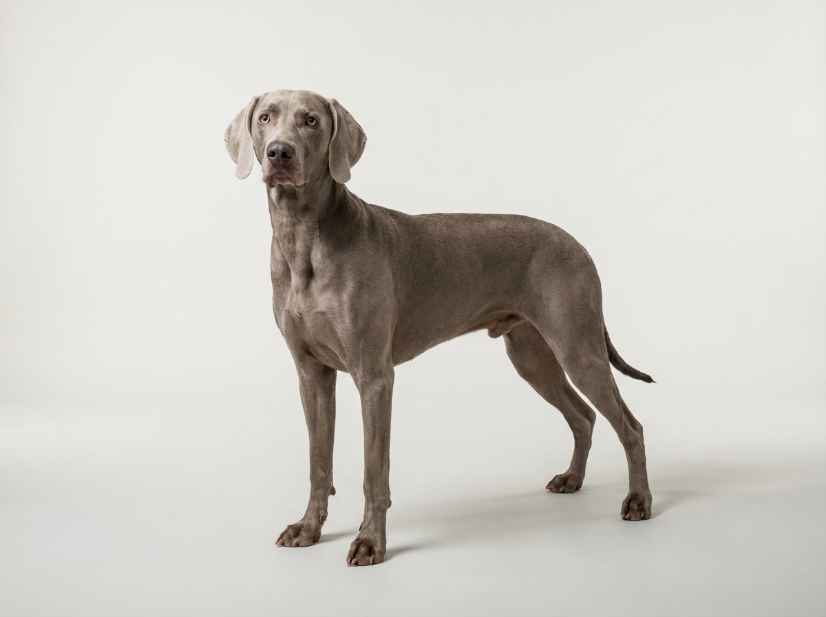
Eighteen-month-old Zola, a Weimaraner belonging to Theo, developed a sudden pink lump in the corner of her left eye one morning that hadn't been there the night before. Theo hadn't heard of cherry eye in Weimaraners specifically and worried it might be something more serious, but his vet identified it immediately as a classic third eyelid gland prolapse. Zola had outpatient surgery within the week to reposition the gland, wore an E-collar for ten days afterward, and healed without complication. Her other eye never developed the same issue.
Key takeaway: Cherry eye isn't a condition Weimaraner owners are typically warned to expect, which is exactly why Theo didn't recognize it right away. It's uncommon in this breed, but a vet can identify and fix it quickly regardless.
No, it isn't documented as a hallmark issue for this breed the way it is for some others with different facial conformation. It can still occur occasionally, since the underlying cause isn't exclusive to any one breed's build.
It's not typically an emergency in the sense of immediate sight loss, but it should be seen by a vet promptly, since the exposed gland can become irritated, dry, or infected the longer it's left untreated.
Occasionally a mild prolapse resolves briefly on its own, but it commonly recurs, and most cases need veterinary treatment, whether that's temporary management or surgical repositioning, to resolve properly.
Removing the gland was an older approach that's now avoided in most cases because that gland produces a significant portion of the eye's tear film; removing it raises the risk of chronic dry eye later in life. Surgical repositioning preserves the gland's function.
A veterinary ophthalmologist or experienced general vet repositions and tacks the gland back into its normal location under anesthesia, an outpatient procedure with a relatively quick recovery.
It varies by region and whether a specialist is involved, generally running from several hundred to over a thousand dollars depending on complexity and whether both eyes are affected.
Yes, it can occur in one eye first and later in the other, though this isn't universal. Each eye is generally treated as its own case if it happens.
No, it's not a documented hallmark issue for the breed, though it can still occur occasionally in any dog.
A pink or reddish, rounded mass appearing suddenly in the inner corner of one or both eyes.
Most cases need surgical repositioning rather than resolving on their own, since prolapse commonly recurs without treatment.
It's avoided in most modern practice because the gland supports tear production; repositioning it instead preserves eye health long term.
Generally several hundred to over a thousand dollars, depending on region, complexity, and whether one or both eyes are affected.
A one-page prep sheet for this condition: the signs, the questions to ask, what to get priced. Unlocks here.
You get: Vet visit prep sheet
No spam. One-click unsubscribe. See our privacy policy.
See a sample page before you decide.
Early access list
The free prep sheet covers the same-day warning signs, a symptom log and the questions to ask. We are considering a fuller kit that adds a cost worksheet for the usual tests and treatments (US price ranges with their sources) and a plain-English guide to ways of covering the bill. It is not built yet; this list tells us whether it is worth building.
One email, no spam, unsubscribe anytime. Information only, not veterinary advice.