Health
Is Legg-Calve-Perthes common in Shelties?
No. It is a toy and small terrier breed condition, and a Sheltie sits at the upper edge of the affected size range. It is possible but uncommon.

Quick answer
Is Legg-Calve-Perthes common in Shelties?
No. It is a toy and small terrier breed condition, and a Sheltie sits at the upper edge of the affected size range. It is possible but uncommon.
It is a disease of small breeds and of puppies, and the entire diagnostic window sits between about four and twelve months of age.
Owners of a Sheltie puppy or adolescent with a limp on one back leg that has not resolved with rest, and who have been told to wait and see for longer than feels right.
This guide is not medical advice. If your dog shows pain, sudden behavior change, or worsening symptoms, consult a licensed veterinarian.
Some links may be affiliate links. We may earn a commission at no extra cost to you. We only suggest products we believe are helpful for dog owners.
We research and check what we publish, but we do not warrant that any figure, date, price, or statement is accurate, complete, or current, and we accept no liability for any loss, cost, injury, or other harm arising from the use of this site. Use it as one reference among several. You remain responsible for your own decisions and for your dog's care - confirm anything that matters with a licensed veterinarian or another qualified professional before you act on it. Full disclaimer.
Legg-Calve-Perthes disease is the loss of blood supply to the femoral head in a growing small-breed dog, causing the bone to die, collapse and deform. It presents as a progressively worsening lameness on one back leg in a puppy between about four and twelve months, with visible muscle wastage on the affected side within weeks. It is uncommon in Shetland Sheepdogs but sits on the differential for any adolescent Sheltie with a limp that will not resolve.
Rare in this breed. Legg-Calve-Perthes is concentrated in toy breeds and small terriers, and a Shetland Sheepdog is larger than the typical patient. Most persistent hind limb lameness in a young Sheltie turns out to be a kneecap or a soft tissue injury rather than this.
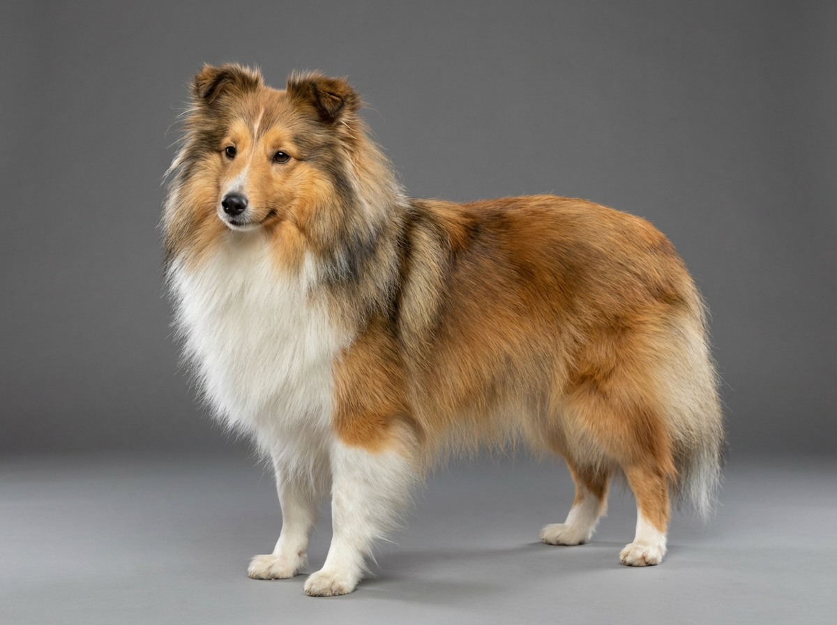
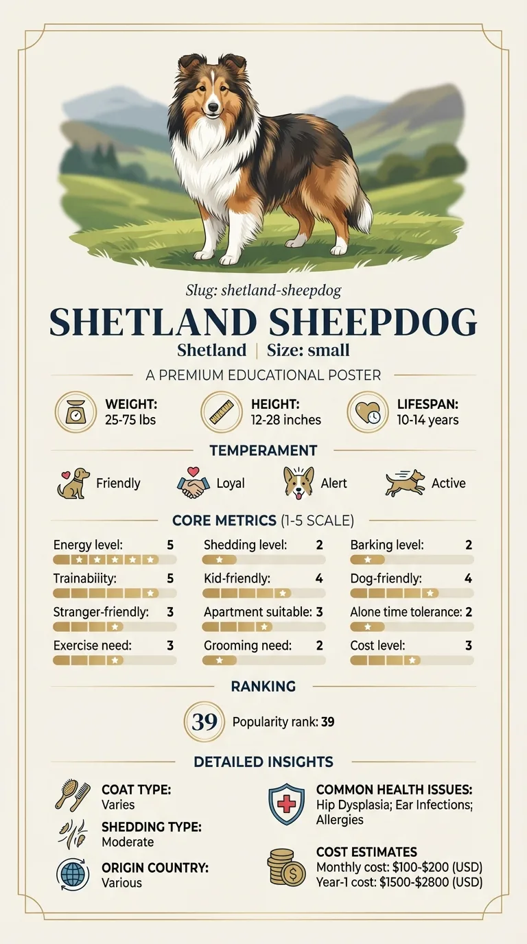
Body size is the connection, and it is a weak one for this breed. At fifteen to twenty five pounds a Sheltie sits above the toy range where the disease clusters, and it is absent from the breed's documented health list. What is genuinely breed-relevant is what surrounds the diagnosis rather than the disease itself: patellar luxation is a documented Sheltie condition and competes for the same clinical picture, and MDR1 drug sensitivity governs the sedation required for the radiographs that settle the question in an adolescent dog.
Nothing an owner does causes this, and it is worth saying so plainly, because parents of affected puppies often search for a fall or a game to blame. What environment can influence is the aftermath. A puppy allowed to keep loading a collapsing femoral head deforms it further, so restricting activity once lameness appears is genuinely useful even before a diagnosis exists. Slippery flooring and repeated jumping down from height are worth removing from a limping adolescent's day regardless of the eventual cause.
Book promptly, within days, for any hind limb lameness in a puppy between four and twelve months that has lasted more than a week, and urgently if the dog is refusing to bear weight at all, if the limb is held up constantly, or if the puppy cries when the hip is touched. Non-weight-bearing lameness in a young dog can also mean a fracture and should be seen the same day. Do not accept an open-ended wait and see for a lameness that has already had two weeks of proper rest; ask for radiographs and ask what the plan is if they are normal. Confirm the MDR1 result is recorded before sedation is booked.
See all Shetland Sheepdog health problems, which breeds are prone to legg calv perthes disease, or the full Shetland Sheepdog breed guide for temperament, exercise needs and ownership costs.
From first limp to diagnosis is typically two to six weeks in a dog whose owner pushes for imaging. Surgery is a single procedure with initial recovery over two weeks and a structured rehabilitation programme running two to four months. Full return of muscle and confident use of the leg often takes six months. Dogs treated conservatively follow a longer and less predictable path, and some end up having surgery later anyway.
Success is a young adult Sheltie that runs, jumps and plays without visible discomfort, with thigh muscle that has rebuilt to something close to the other side. A slight difference in gait when the dog is tired is normal and does not represent failure. What you should not accept as an endpoint is a dog that has simply learned to use three legs, which usually means muscle was never rebuilt.
The femoral head in a young dog is fed by a small and rather precarious network of vessels. If that supply is interrupted, the bone tissue dies. Dead bone does not hurt in itself, but it also does not remodel or repair normally, and it cannot bear weight the way living bone does.
Over the following weeks the body begins to clear the dead tissue and lay down new bone. During that process the head is structurally weak, and it deforms. What was a smooth sphere sitting neatly in the hip socket becomes irregular and flattened. The joint surfaces no longer match, and the resulting grinding is painful and progressive.
The pain therefore builds rather than arriving suddenly, which is why owners so often describe a limp that came and went for a few weeks before becoming constant.
Why the blood supply fails in the first place is not fully established. A hereditary component is generally accepted in the small breeds where it is common, and it is typically one-sided, though a minority of dogs are affected in both hips.
This is a toy and small terrier breed disease. Its usual patients are the very small: Yorkshire Terriers, Miniature Pinschers, West Highland Whites, Pugs and similar. A Shetland Sheepdog at fifteen to twenty five pounds sits at the upper edge of the size range where it is reported, and it does not appear on the breed's documented health list, which covers Collie Eye Anomaly, MDR1 drug sensitivity, dermatomyositis, hypothyroidism and patellar luxation.
So it is uncommon here. But an adolescent Sheltie with a persistent one-sided hind limb lameness has a short differential, and this belongs on it alongside two more likely candidates.
The first competitor is patellar luxation, which is a documented Sheltie problem. The classic tell is intermittency: the dog skips for a stride or two, then runs on normally. Perthes disease does not behave that way; it gets steadily worse and the dog uses the leg less and less.
The second is injury, which in a bouncy adolescent herding dog is always plausible and usually improves with a fortnight of restriction. A young Sheltie whose limp has not improved after two weeks of genuine rest has crossed the line from wait and see into radiograph.
Those radiographs need sedation, which is where MDR1 status becomes part of the plan rather than a footnote.

If you think your Shetland has legg-calvé-perthes disease, the plan is three steps: write down what you have seen and when it started, book a veterinary appointment rather than waiting for the next flare, and take video of the behaviour or symptom before you go — the thing you are worried about rarely happens in the consulting room. Screening in the parents covers collie eye anomaly, mdr1 drug sensitivity, dermatomyositis.
What the vet visit should produce is a diagnosis and a written plan, not just reassurance: what is being ruled out, what the monitoring interval is, and which signs mean you come back sooner. Ask what the treatment costs across a year rather than per visit, because that is the number that decides whether you are managing this condition or reacting to it.
Between appointments, keep a short log — dates, what you saw, what changed. It is the single most useful thing an owner brings to a follow-up, and for legg-calvé-perthes disease it is often what separates a clear pattern from a guess.
Sinead's Sheltie puppy, Brodie, started favouring his left hind leg at six months. Her practice advised rest and a recheck, twice, over six weeks. Each time he seemed slightly better after a quiet week and worse again after a weekend. What changed her mind was running a hand down both thighs while grooming him and realising the left one was noticeably thinner. She asked directly for radiographs and for the MDR1 test to be run first, having read that Shelties needed it. The images showed a collapsed and irregular femoral head. Brodie had the head removed at seven and a half months, started physiotherapy at two weeks post-operatively, and was doing hydrotherapy weekly through the spring. At eighteen months he was a normal, obnoxiously energetic adolescent Sheltie. His surgeon noted that the muscle wastage Sinead had felt was the finding that should have triggered imaging a month earlier.
Key takeaway: A limp that improves with rest and returns with activity in a four to twelve month old dog is not resolving, it is cycling. Feel for thigh muscle asymmetry under the coat, and let that, rather than another fortnight of rest, be the trigger for radiographs.
Almost always between four and twelve months, occasionally a little later. It is a disease of the growing skeleton. A hind limb lameness starting in a mature Sheltie is something else and should not be investigated down this path.
On radiographs of the hips, which show a femoral head that has lost its normal density and shape. Because a puppy has to be positioned precisely and held still, this means sedation. Confirm your Sheltie's MDR1 genotype before that appointment rather than during it.
No, though both affect the hip. Dysplasia is a joint that formed with poor congruity between ball and socket, usually affects both sides, and is a large-breed problem. Perthes is the death and collapse of the ball itself due to lost blood supply, is usually one-sided, and is a small-breed puppy problem.
In most cases the surgical option is removal of the damaged femoral head, after which the body forms a fibrous false joint. It sounds drastic and it works remarkably well in small dogs, because there is not much weight to carry. Some very mild cases are managed conservatively with pain control and physiotherapy, but that decision belongs to a surgeon.
Most small dogs return to good function, with an athletic gait that may never be quite symmetrical. The muscle rebuilt after surgery is what determines the result, which is why physiotherapy is not optional. Many go on to normal activity, and some compete in sport at a modest level.
It usually affects one, but a minority of dogs are bilaterally affected, sometimes months apart. If your puppy has had one hip treated, a new lameness on the other side deserves prompt imaging rather than an assumption that lightning does not strike twice.
No. It is a toy and small terrier breed condition, and a Sheltie sits at the upper edge of the affected size range. It is possible but uncommon.
Two weeks of genuine restriction. A hind limb lameness in a four to twelve month old dog that has not improved by then needs radiographs.
Perthes tends to worsen steadily. An intermittent skip that resolves within a stride or two points more toward the kneecap, which is a documented Sheltie issue.
Femoral head removal in a small dog commonly runs into the low thousands including imaging and follow-up, well beyond routine ownership costs.
Yes. The false joint that forms is supported entirely by muscle, so the rehabilitation determines the outcome more than the surgery does.
A one-page prep sheet for this condition: the signs, the questions to ask, what to get priced. Unlocks here.
You get: Vet visit prep sheet
No spam. One-click unsubscribe. See our privacy policy.
See a sample page before you decide.
Early access list
The free prep sheet covers the same-day warning signs, a symptom log and the questions to ask. We are considering a fuller kit that adds a cost worksheet for the usual tests and treatments (US price ranges with their sources) and a plain-English guide to ways of covering the bill. It is not built yet; this list tells us whether it is worth building.
One email, no spam, unsubscribe anytime. Information only, not veterinary advice.