Health
What is the Sheltie's main inherited eye condition?
Collie Eye Anomaly, a developmental fault in the vascular layer beneath the retina. It is documented on the breed's health profile.

Quick answer
What is the Sheltie's main inherited eye condition?
Collie Eye Anomaly, a developmental fault in the vascular layer beneath the retina. It is documented on the breed's health profile.
There is a window of a few weeks in a Sheltie puppy's life when an inherited eye condition can be seen clearly, and after which developing pigment can hide it. Missing that window is why so many adult dogs have an uncertain eye status.
Prospective owners trying to understand what eye documentation from a breeder actually means. People who have just been told their dog is affected and want to know what happens next. Anyone considering breeding and needing to understand carrier status.
This guide is not medical advice. If your dog shows pain, sudden behavior change, or worsening symptoms, consult a licensed veterinarian.
Some links may be affiliate links. We may earn a commission at no extra cost to you. We only suggest products we believe are helpful for dog owners.
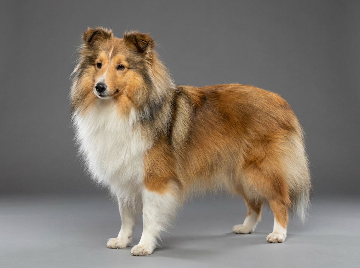
Collie Eye Anomaly is an inherited developmental abnormality of the tissue layer that supplies blood to the retina. It is present at birth, it does not develop later, and it spans a wide range of severity from a thin patch with no functional consequence to structural defects that lead to retinal detachment and blindness. It is documented for the Shetland Sheepdog and shared with the Rough Collie and other related herding breeds through common ancestry.
CEA is one of only five conditions documented for this breed, which places it among the most relevant inherited concerns a Sheltie owner has. The carrier rate in the breed is high enough that responsible breeders test every breeding animal as standard practice. Most affected dogs sit at the mild end and live entirely normal visual lives, which is why the condition persists: it is often invisible without a specialist looking for it.
This is a purely genetic condition traceable to the Shetland Sheepdog's shared ancestry with the Rough Collie, in which the same variant is well established. It is inherited in a recessive pattern, meaning a dog needs two copies to be affected and carriers with one copy are entirely normal. That is precisely why it has persisted through generations of selection: carriers show nothing, so without DNA testing the variant travels silently. Nothing about the breed's coat, size or head shape contributes. The eye's almond shape and well-set orbit are unrelated to the choroid's development.
Nothing in the environment causes CEA, and no diet, supplement or management change affects whether a puppy is born with it. Where environment matters is at the severe end, since an eye with a large coloboma is structurally weaker and a blow to the head can precipitate a retinal detachment in a way it would not in a normal eye. Bright sunlight and outdoor work do not cause the condition but a dog with reduced vision navigates less confidently in low light, which shapes practical management.
Seek same-day care for sudden apparent vision loss, for a dilated pupil that does not respond to light, for a dog suddenly bumping into things, or for an eye that is painful, red or being held shut. Retinal detachment causes sudden loss of vision in the affected eye and needs urgent specialist assessment. Book a specialist ophthalmology examination for any puppy from an untested line at six to eight weeks, since the window closes as pigment develops. For an adult dog of unknown status, ask about DNA testing, which is unaffected by age and gives an answer the eye examination may no longer provide.
See all Shetland Sheepdog health problems, which breeds are prone to eye disorders, or the full Shetland Sheepdog breed guide for temperament, exercise needs and ownership costs.
Diagnosis happens in a single specialist appointment, ideally at six to eight weeks. DNA results take two to three weeks. There is no treatment timeline because the malformation itself is not treatable, and mildly affected dogs need nothing beyond periodic checks across a 12 to 14 year life.
For most affected Shelties, success is a documented diagnosis and a dog who sees perfectly well for life. For severely affected ones it is early recognition, annual monitoring, and prompt action if a detachment occurs. At the breed level success is testing every breeding animal so that affected puppies stop being produced.
CEA is not one thing. It spans a range of severity, and the practical implications differ enormously between the ends of it.
There are two ways to assess CEA and they answer different questions. A specialist ophthalmologist examining a litter at six to eight weeks looks at what the eye actually is, and that examination detects the physical changes including the severe forms. A DNA test looks at whether the dog carries the associated genetic variant, and it returns clear, carrier or affected.
Both have limits. The eye examination has the go normal problem: mild lesions can be masked by pigment developing after about eight weeks, so a dog examined at six months may appear normal when she is not. That is why litter screening happens when it does and cannot simply be deferred. The DNA test avoids that timing problem entirely, but it identifies the genetic status rather than the actual severity, and it does not predict which affected dogs will have colobomas or detachments.
What that means in practice is that a responsible Shetland Sheepdog breeder does both: DNA tests the parents so pairings can be planned, and has the litter examined by an ophthalmologist within the window. As a buyer, ask for both in writing. A verbal reassurance that the line has never had eye problems is not documentation, and lines can carry the variant silently for generations because carriers are unaffected.
For a dog already diagnosed, the honest position is that there is no treatment for the underlying malformation. Mildly affected dogs need nothing beyond periodic ophthalmic checks. Dogs with significant colobomas are monitored for detachment. What a diagnosis genuinely changes is breeding decisions and the baseline against which any future eye problem is judged, which is more useful than it sounds when a dog turns up squinting at six years old.

If you think your Shetland has eye disorders, the plan is three steps: write down what you have seen and when it started, book a veterinary appointment rather than waiting for the next flare, and take video of the behaviour or symptom before you go — the thing you are worried about rarely happens in the consulting room. Screening in the parents covers collie eye anomaly, mdr1 drug sensitivity, dermatomyositis.
What the vet visit should produce is a diagnosis and a written plan, not just reassurance: what is being ruled out, what the monitoring interval is, and which signs mean you come back sooner. Ask what the treatment costs across a year rather than per visit, because that is the number that decides whether you are managing this condition or reacting to it.
Between appointments, keep a short log — dates, what you saw, what changed. It is the single most useful thing an owner brings to a follow-up, and for eye disorders it is often what separates a clear pattern from a guess.
Elspeth had bred Shelties for eight years and DNA tested every breeding dog. A carrier-to-clear pairing should not produce affected puppies, and it did not, but she booked the ophthalmologist for the litter at seven weeks anyway because her breed club expected it. All six puppies examined normal. The value of the certificate showed up two years later, when one of those puppies, by then called Bracken, began bumping into things in the garden at dusk. Her owner's vet had the seven week certificate on file and could say confidently that this was new rather than inherited. The investigation found a different problem entirely, unrelated to CEA, and it was caught early because nobody wasted a month wondering whether the eye had always been like that.
Key takeaway: The screening certificate is worth having even when it is normal. A documented baseline is what makes a later change unambiguous.
Most affected dogs will not. The mild form, choroidal hypoplasia, typically causes no functional vision loss at all. Blindness is associated with the severe end of the spectrum, particularly large colobomas and retinal detachment, which are a minority of cases.
The underlying malformation is developmental and does not progress. What can happen later is a complication such as retinal detachment in a severely affected eye, which is why dogs with significant lesions are monitored rather than discharged.
It describes mild lesions becoming obscured by pigment that develops in the eye after around eight weeks, so an affected dog can examine as normal later in life. It is the single reason litter screening must happen at six to eight weeks rather than whenever is convenient.
With care and with genetic advice. Carriers have one copy of the variant and are themselves unaffected. Breeding two carriers together is what produces affected puppies, which is why DNA testing both parents allows pairings to be planned rather than gambled on.
Written DNA results for both parents and a specialist ophthalmologist's litter examination certificate. Ask for copies rather than descriptions, and note that this breed's other worthwhile test is MDR1, which is equally documented and equally important.
Bumping into furniture, hesitancy in dim light, a dilated pupil that does not respond to light, or a dog who suddenly becomes clingy and cautious. Sudden apparent vision loss deserves a same-day appointment, whatever the underlying cause.
Collie Eye Anomaly, a developmental fault in the vascular layer beneath the retina. It is documented on the breed's health profile.
At six to eight weeks by a specialist ophthalmologist, before developing pigment can mask mild lesions.
No. Most affected dogs have the mild form and see normally. Blindness is confined to the severe end of the spectrum.
Written DNA results for both parents plus a specialist litter eye certificate. Also ask about MDR1 testing.
A specialist examination and a DNA test are modest one-off costs against the breed's eighty to a hundred and fifty dollar monthly running expenses.
A one-page prep sheet for this condition: the signs, the questions to ask, what to get priced. Unlocks here.
You get: Vet visit prep sheet
No spam. One-click unsubscribe. See our privacy policy.
Preview page 1 before you decide.