Health
What is the single most urgent sign of glaucoma?
A red, painful eye with a cloudy cornea and a poorly responsive pupil. This combination needs same-day emergency veterinary care.

Quick answer
What is the single most urgent sign of glaucoma?
A red, painful eye with a cloudy cornea and a poorly responsive pupil. This combination needs same-day emergency veterinary care.
Glaucoma is not a description of a red eye. It is a specific, measurable rise in pressure inside the eyeball, and in this breed it is usually a consequence of something else going wrong first, not a stand-alone disease waiting to appear.
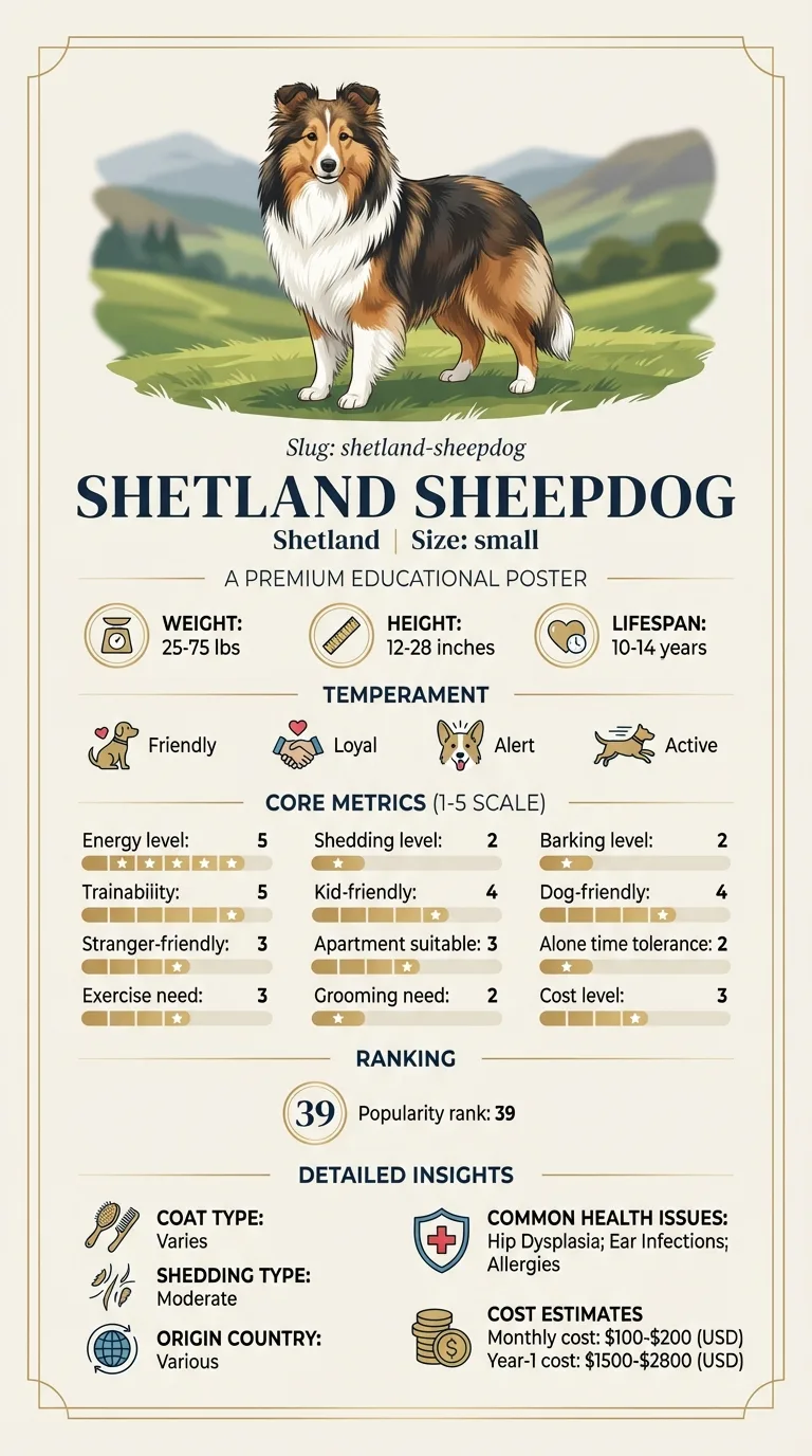
Owners of a Sheltie with a sudden red, painful, watering eye who need to know how urgently to act. People whose dog has a known Collie Eye Anomaly finding and want to understand what raises their specific risk. Anyone who has already had one eye affected and wants to understand what happens to the second.
This guide is not medical advice. If your dog shows pain, sudden behavior change, or worsening symptoms, consult a licensed veterinarian.
Some links may be affiliate links. We may earn a commission at no extra cost to you. We only suggest products we believe are helpful for dog owners.
We research and check what we publish, but we do not warrant that any figure, date, price, or statement is accurate, complete, or current, and we accept no liability for any loss, cost, injury, or other harm arising from the use of this site. Use it as one reference among several. You remain responsible for your own decisions and for your dog's care - confirm anything that matters with a licensed veterinarian or another qualified professional before you act on it. Full disclaimer.
Glaucoma is a rise in internal eye pressure that compresses the optic nerve and can cause permanent vision loss within one to two days if untreated. It is not one of the Shetland Sheepdog's documented conditions in the primary, inherited form seen in some other breeds. The relevant risk in Shelties is secondary glaucoma, arising as a complication of significant Collie Eye Anomaly findings such as colobomas or retinal detachment, rather than a stand-alone drainage-angle defect.
Primary glaucoma is uncommon to rare in this breed and is not documented as a Shetland Sheepdog predisposition. Secondary glaucoma is less common than the underlying conditions that cause it, but it is a genuine and serious risk specifically in the minority of Shelties with significant colobomas or a history of retinal detachment, which is why those dogs warrant closer monitoring than the breed average.
The Shetland Sheepdog's relevant glaucoma risk is inherited indirectly, through its documented predisposition to Collie Eye Anomaly rather than through any drainage-angle defect of its own. A coloboma or chronic retinal detachment changes the internal environment of the eye in ways that can obstruct normal fluid outflow over time, and it is this secondary pathway, not a primary glaucoma gene, that explains why glaucoma appears in this breed at all. Dogs with only mild choroidal hypoplasia, the most common and functionally harmless form of CEA, do not carry this elevated risk.
There is no dietary, environmental or lifestyle trigger for glaucoma itself. What does matter is monitoring: an eye with a known coloboma benefits from periodic pressure checks by an ophthalmologist so early elevation is caught before vision is threatened, and any blunt trauma to a structurally vulnerable eye should be taken seriously given the underlying fragility a coloboma creates. Delayed recognition of a red, painful eye, rather than any environmental cause, is the main modifiable factor in outcome.
Treat as a true emergency, requiring same-day assessment without delay: a red, painful eye with a cloudy or bluish cornea, a dilated pupil that responds poorly to light, sudden apparent vision loss, or an eye that looks larger than its fellow. Every hour of delay in confirmed acute glaucoma is a real risk to remaining vision. Book promptly, though not necessarily same-day, for a known coloboma or retinal detachment history due a routine pressure check, or for any new redness or discomfort in a dog with that history. Mention MDR1 status before any sedation or eye surgery is planned.
See all Shetland Sheepdog health problems, which breeds are prone to glaucoma, or the full Shetland Sheepdog breed guide for temperament, exercise needs and ownership costs.
Acute glaucoma is diagnosed in a single emergency visit using tonometry, and treatment to lower pressure begins immediately once confirmed. Vision already lost to sustained high pressure does not return, so the treatment timeline afterwards is about preserving whatever vision and comfort remain, often for the rest of the dog's life, with periodic pressure checks continuing indefinitely.
For a dog caught early, success is stable eye pressure on ongoing medication and preserved vision in that eye. For a dog diagnosed later, success is a comfortable, pain-free eye, whether sighted or not, and close monitoring of the fellow eye to catch any early change before it progresses to the same point.
Glaucoma can develop gradually in some cases, but the presentation that matters most to recognise is the acute one, because vision lost to sustained high pressure is not recoverable and the window to save it is measured in hours, not days.
Primary glaucoma, an inherited structural fault in the eye's drainage angle present from early life, is well documented in breeds such as Cocker Spaniels and Basset Hounds. It is not part of the Shetland Sheepdog's documented health profile, and a Sheltie developing sudden high eye pressure should not automatically be assumed to have that form.
What is mechanistically relevant to this breed is secondary glaucoma, meaning pressure elevation caused by another problem interfering with normal drainage. In Shelties, the most breed-relevant route runs through Collie Eye Anomaly. A coloboma, a structural gap in the tissue at the back of the eye, can predispose to retinal detachment, and a detached retina releases inflammatory material and disrupts the eye's internal architecture in ways that can obstruct fluid drainage and raise pressure. Chronic uveitis, inflammation inside the eye from any cause, is another well recognised route to secondary glaucoma, as can be a displaced or cataract-affected lens blocking normal fluid flow.
This distinction matters practically. A dog with a known coloboma or a history of retinal detachment carries a genuinely elevated risk of secondary glaucoma in that eye and deserves closer monitoring, including a specific discussion with an ophthalmologist about pressure checks, rather than being told glaucoma is simply unlikely for the breed in general. A dog with a normal eye examination and no CEA findings, by contrast, has no particular breed-specific reason to expect glaucoma at all.
One further practical point. Glaucoma in one eye significantly raises the risk that the second eye will eventually be affected too, whatever the underlying cause, because whatever process disrupted drainage in the first eye is often relevant to the anatomy of the second. Once one eye has been diagnosed, a vet will usually recommend monitoring, and sometimes preventive treatment, for the fellow eye even while it still looks and functions normally.

Reuben was a Sheltie diagnosed at eight weeks with a moderate coloboma in his left eye, noted on his breeder's certificate but causing no visible problem through his first six years. His owner, Marcus, had been told the finding was there and to mention it at vet visits, which he dutifully did, without it ever becoming more than a line in the notes. At seven, Reuben's left eye turned red overnight and he began pawing at his face. Marcus, remembering the old certificate, went straight to an emergency vet rather than waiting to see if it settled. Tonometry confirmed markedly elevated pressure. The retina in that eye had detached, triggering secondary glaucoma. Treatment controlled the pressure and Reuben kept the eye, though vision in it did not return. His right eye, previously never flagged as a concern, is now checked every six months given his history, and remains completely normal three years on.
Key takeaway: A coloboma that causes no problem for years is not the same as a coloboma that will never cause one. Knowing the history is what got Reuben to the right kind of urgent care instead of a wait-and-see night at home.
Not as a primary, inherited condition, which is not documented for this breed. The relevant risk is secondary glaucoma arising from complications of Collie Eye Anomaly, particularly in a dog with a known coloboma or history of retinal detachment.
With a handheld instrument called a tonometer, which gently touches the surface of a numbed eye and gives a pressure reading in seconds. It is quick, well tolerated, and the only reliable way to confirm glaucoma rather than guessing from appearance.
In acute, severely elevated pressure, permanent vision loss can occur within twenty four to forty eight hours, sometimes faster. This is why a red, painful eye with a cloudy cornea should be treated as same-day, not next-week, veterinary care.
Generally, yes. The elevated secondary glaucoma risk is specifically associated with more significant findings such as colobomas and retinal detachment, not with the common mild form, choroidal hypoplasia, which typically causes no functional consequence at all.
Underlying pressure can often be controlled with medication, and some cases need surgery, but a glaucoma episode that has already damaged the optic nerve does not restore lost vision. The goal of treatment is preserving remaining vision and comfort, not reversing what has already happened.
In advanced, unrelenting cases where pressure cannot be controlled and the eye is permanently blind and painful, removal of the eye is sometimes the kindest option. Dogs adapt remarkably well to vision in one eye or none at all, and pain relief is the priority once sight cannot be recovered.
Yes, routinely. The fellow eye carries meaningfully elevated risk once one eye is affected, and your vet will likely recommend regular pressure checks and sometimes preventive treatment for it even while it still appears normal.
A red, painful eye with a cloudy cornea and a poorly responsive pupil. This combination needs same-day emergency veterinary care.
No, primary glaucoma is not documented in this breed. Risk here runs through complications of Collie Eye Anomaly rather than a stand-alone inherited drainage fault.
With a tonometer, a handheld instrument that measures internal eye pressure directly on a numbed eye in seconds.
Emergency pressure-lowering treatment, ongoing medication and specialist monitoring commonly runs into hundreds to low thousands of dollars, well beyond the breed's typical monthly costs.
Yes, remarkably well. This breed's intelligence and trainability make adapting cues and household layout for reduced vision genuinely effective.
A one-page prep sheet for this condition: the signs, the questions to ask, what to get priced. Unlocks here.
You get: Vet visit prep sheet
No spam. One-click unsubscribe. See our privacy policy.
Preview page 1 before you decide.