Health
What is corneal dystrophy?
An inherited, non-inflammatory deposit of lipid or mineral material in the cornea, producing a painless, usually symmetrical white or grey opacity on the eye's surface.

Quick answer
What is corneal dystrophy?
An inherited, non-inflammatory deposit of lipid or mineral material in the cornea, producing a painless, usually symmetrical white or grey opacity on the eye's surface.
A hazy, greyish-white patch appears on the clear surface of the eye — often oval, often centred, often mirrored almost exactly on the other side. The dog is not squinting. The dog is not rubbing. The dog is behaving completely normally and appears to see fine. That combination of a visible opacity with a comfortable dog is the signature of corneal dystrophy, and it separates this from almost everything else that clouds an eye.
Owners who spot a cloudy eye and reasonably fear the worst — cataracts, an ulcer, an injury, going blind. Also owners of short-coated breeds where nothing obscures the face, so the change is noticed immediately and creates alarm out of proportion to what is usually a manageable finding.
This guide is not medical advice. If your dog shows pain, sudden behavior change, or worsening symptoms, consult a licensed veterinarian.
Some links may be affiliate links. We may earn a commission at no extra cost to you. We only suggest products we believe are helpful for dog owners.
We research and check what we publish, but we do not warrant that any figure, date, price, or statement is accurate, complete, or current, and we accept no liability for any loss, cost, injury, or other harm arising from the use of this site. Use it as one reference among several. You remain responsible for your own decisions and for your dog's care - confirm anything that matters with a licensed veterinarian or another qualified professional before you act on it. Full disclaimer.
An inherited, non-inflammatory deposit of lipid or mineral material within the cornea, producing a symmetrical, painless white or grey opacity. Most cases require monitoring rather than treatment. It becomes clinically significant if it interferes with vision or if the corneal surface ulcerates over a deposit.
Corneal dystrophies are recognised across many breeds and are among the more frequent causes of a chronic, non-painful corneal opacity. In this breed, corneal dystrophy is a documented consideration, and it sits apart from the breed's inflammatory and orthopaedic problems because nothing about lifestyle, activity or environment brings it on.
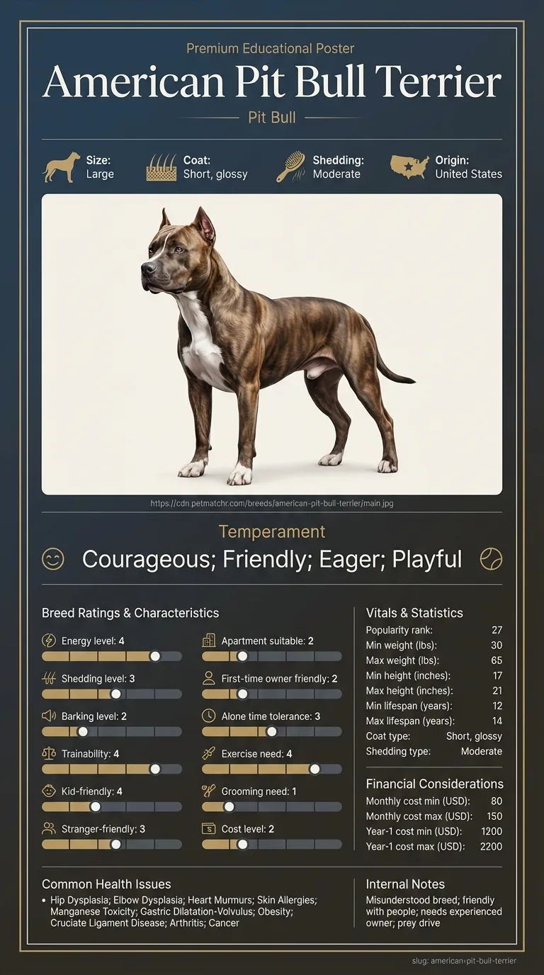
Corneal dystrophy is documented as a consideration in this breed, and the mechanism is genetic rather than structural — nothing about a 30-to-65-pound athletic build predisposes the cornea to deposit material. What the breed's short, glossy coat and open facial structure do contribute is visibility: there is no coat obscuring the eye, so owners notice an opacity early, which is genuinely helpful for distinguishing a stable dystrophy from a progressive problem. The breed's documented hypothyroidism-adjacent endocrine considerations are worth mentioning to a vet, since thyroid status can be relevant to corneal lipid deposition.
None cause it. Environment matters only for the complications: dust, wind and rough face-first play raise the risk of corneal trauma, and trauma over an existing deposit is the route by which a painless dystrophy becomes a painful ulcer. Untreated allergic itching that drives face rubbing works the same way.
Book an appointment for any new corneal opacity, even a painless one, so it can be stained and characterised properly — distinguishing a dystrophy from an ulcer is a two-minute in-clinic test and it changes everything about what happens next. Ask whether bloodwork is warranted to check thyroid function and lipid levels, since corneal deposits can reflect a systemic issue. Seek same-day attention if the eye becomes painful, if the dog squints or holds it closed, if there is discharge, redness or visible surface disruption, or if vision appears to change — that suggests ulceration or a separate problem, and painful eye conditions deteriorate quickly. Ongoing, a stable dystrophy is usually rechecked at routine appointments rather than needing its own schedule.
Start here: American Pit Bull Terrier Most Common Genetic Disorders
See all American PIT Bull Terrier health problems, which breeds are prone to corneal dystrophy, or the full American PIT Bull Terrier breed guide for temperament, exercise needs and ownership costs.
Most dystrophies are stable or change very slowly over years. There is no treatment that dissolves the deposits, and no eye drop that clears them — that is worth stating plainly, because it is the first thing owners look for. Monitoring is the management. If ulceration occurs over a deposit, that episode is treated on its own timeline, usually a couple of weeks of medication and rechecks.
Success here is usually uneventful: an opacity that stays roughly the same, a dog that continues to see and behave normally, and an owner who has stopped worrying about it because it has been properly identified. Where an underlying thyroid or lipid problem is found, success includes treating that — which occasionally improves the corneal picture but matters far more for the dog's general health than for the eye.
Owners cannot reliably diagnose this at home, but knowing the categories makes for a far more useful vet visit.
A corneal ulcer is painful. The dog squints, holds the eye shut, tears excessively and often paws at its face. It is typically one eye, and it is urgent — ulcers can deteriorate fast and threaten the eye.
A cataract sits deeper, in the lens rather than on the surface, and looks like clouding behind the pupil rather than a patch on the front. It affects vision progressively.
Nuclear sclerosis is the bluish haze of an ageing lens, normal in older dogs and not vision-threatening.
Corneal dystrophy sits in the cornea itself, on the surface, and the dog is comfortable. Symmetrical, painless and non-progressive-to-slowly-progressive is the pattern. A vet distinguishes these directly — a fluorescein stain will light up an ulcer immediately and will not stain a dystrophy — which is exactly why a cloudy eye is worth an appointment rather than a search engine.
Most corneal dystrophy in dogs is a cosmetic finding that never needs treatment. The deposits sit in the cornea, vision is largely unaffected because the opacity rarely covers enough of the visual axis to matter, and the correct management is periodic monitoring.
There are two circumstances where that changes. The first is if deposits become dense or extensive enough to genuinely interfere with vision, which is uncommon. The second, and more clinically relevant, is if the corneal surface breaks down over a deposit and ulcerates — at which point a painless condition becomes a painful one and needs treating as an ulcer.
There is also a version worth flagging: corneal deposits can sometimes reflect an underlying systemic problem with lipid metabolism, such as hypothyroidism or a lipid disorder. That is why a vet may run bloodwork rather than simply noting the opacity and moving on. It is not routine alarmism — it is checking whether the eye is telling you something about the rest of the dog.

If you think your Pit Bull has corneal dystrophy, the plan is three steps: write down what you have seen and when it started, book a veterinary appointment rather than waiting for the next flare, and take video of the behaviour or symptom before you go — the thing you are worried about rarely happens in the consulting room. Screening in the parents covers hip dysplasia, elbow dysplasia, heart murmurs.
What the vet visit should produce is a diagnosis and a written plan, not just reassurance: what is being ruled out, what the monitoring interval is, and which signs mean you come back sooner. Ask what the treatment costs across a year rather than per visit, because that is the number that decides whether you are managing this condition or reacting to it.
Between appointments, keep a short log — dates, what you saw, what changed. It is the single most useful thing an owner brings to a follow-up, and for corneal dystrophy it is often what separates a clear pattern from a guess.
A four-year-old Pit Bull came in because his owner had noticed a greyish oval patch on his right eye and had spent a bad weekend reading about blindness. In the consulting room two things resolved it quickly. First, the left eye had a nearly identical patch in nearly the same position, which his owner had not registered because she had been fixated on the right. Second, fluorescein stain lit up nothing at all — no ulcer. The vet ran a thyroid panel as a precaution, which came back normal, and recommended photographs every few months. Two years on the patches look the same as the day they were found, and he has never once squinted.
Key takeaway: Symmetry and comfort are the two features that separate this from the eye problems that genuinely need urgent attention.
No, and that is one of its defining features. A cloudy eye in a comfortable, normally behaving dog points away from an ulcer. Pain, squinting or pawing means something else and needs same-day attention.
Usually not. The deposits rarely cover enough of the visual axis to significantly impair sight. Substantial vision loss from corneal dystrophy alone is uncommon.
Location. A cataract is in the lens, seen as clouding behind the pupil. A dystrophy is in the cornea itself, on the front surface of the eye.
Because the cause is inherited rather than acquired. Symmetry is a strong clue pointing to dystrophy rather than to injury or infection, which usually affect one eye.
No. Nothing dissolves the deposits. Management is monitoring, and treating any complications or underlying systemic cause that is identified.
Because corneal lipid deposits can reflect an underlying metabolic issue such as hypothyroidism or a lipid disorder. Checking is sensible medicine, not overkill.
Not entirely, but face-first rough play and dusty, windy conditions raise the risk of corneal trauma — and trauma over an existing deposit is the main route to a painful complication.
The early signs are set out in the sections above, and they are the reason to book a veterinary appointment rather than wait. Corneal Dystrophy in Pit Bulls is diagnosed by a vet, not from a symptom list, and the sooner it is on record the more options remain open.
The early signs are set out in the sections above, and they are the reason to book a veterinary appointment rather than wait. Corneal Dystrophy in American Pit Bull Terriers is diagnosed by a vet, not from a symptom list, and the sooner it is on record the more options remain open.
An inherited, non-inflammatory deposit of lipid or mineral material in the cornea, producing a painless, usually symmetrical white or grey opacity on the eye's surface.
You don't, at home — but ulcers hurt. A comfortable dog with matching opacities in both eyes suggests dystrophy; squinting and pain suggest an ulcer needing same-day care.
Most cases need monitoring rather than treatment. Nothing dissolves the deposits. Treatment is reserved for complications such as ulceration over a deposit.
Corneal deposits can reflect an underlying lipid disorder or thyroid problem, so checking systemic causes is appropriate rather than excessive.
Usually yes. The opacity rarely covers enough of the visual axis to meaningfully affect sight, and most affected dogs behave completely normally.
A one-page prep sheet for this condition: the signs, the questions to ask, what to get priced. Unlocks here.
You get: Vet visit prep sheet
No spam. One-click unsubscribe. See our privacy policy.
Preview page 1 before you decide.